Why Advanced Diagnostics Matter
Early detection of ocular disease is the cornerstone of preserving vision, because many eye conditions—glaucoma, diabetic retinopathy, macular degeneration, and keratoconus—can progress silently before symptoms appear. Optical Coherence Tomography (OCT) provides micrometer‑resolution, cross‑sectional images of retinal layers and the optic nerve head, allowing clinicians to spot subtle thinning or fluid buildup long before visual‑field loss is measurable. Corneal topography creates three‑dimensional curvature maps that reveal irregular astigmatism, early ectasia, and corneal scarring, guiding precise contact‑lens fitting and safe refractive‑surgery planning. Apple Eye Care integrates the DRI OCT Triton and state‑of‑the‑art topography into every comprehensive exam, ensuring that each patient receives technology‑driven, personalized care that translates to earlier intervention, better treatment outcomes, and sustained visual health.
Understanding OCT: The Retina’s 3‑D Blueprint
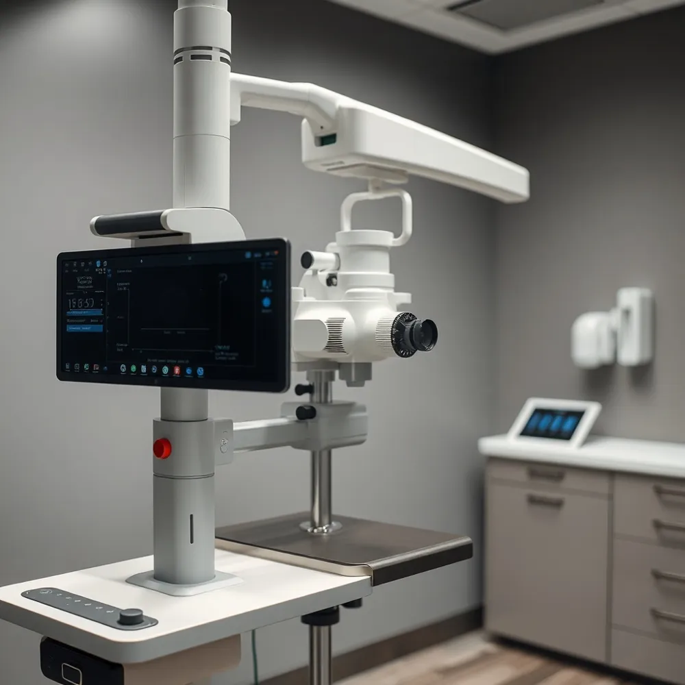
![]() Optical Coherence Tomography (OCT) uses invisible infrared light and low‑coherence interferometry to build high‑resolution, three‑dimensional cross‑sectional images of the retina, macula, optic nerve head, and, in swept‑source systems like the DRI OCT Triton, the anterior segment. By measuring the thickness of each retinal layer, OCT can detect the earliest structural changes of macular degeneration, diabetic retinopathy, and glaucoma—often before symptoms arise—allowing timely treatment and precise monitoring of disease progression.
Optical Coherence Tomography (OCT) uses invisible infrared light and low‑coherence interferometry to build high‑resolution, three‑dimensional cross‑sectional images of the retina, macula, optic nerve head, and, in swept‑source systems like the DRI OCT Triton, the anterior segment. By measuring the thickness of each retinal layer, OCT can detect the earliest structural changes of macular degeneration, diabetic retinopathy, and glaucoma—often before symptoms arise—allowing timely treatment and precise monitoring of disease progression.
During a scan the patient rests a chin on a support, looks at a fixed target, and the machine captures the eye in seconds without any contact. No eye drops are usually needed; only if a clearer view is required may the clinician dil the the pupils, causing short‑term light sensitivity or blurry vision.
The test is painless, takes only a few minutes, and produces digital images that can be stored and compared over time. Apple Eye Care in El Paso incorporates OCT into routine comprehensive exams, providing a non‑invasive, reliable roadmap for preserving vision.
Corneal Topography: Mapping the Front Surface of the Eye
![]() Corneal topography uses either Placido‑disk or Scheimpflug imaging to create a three‑dimensional, color‑coded map of the corneal surface. The Placido‑disk projects concentric rings onto the tear film; reflected mires are analyzed to calculate anterior curvature, while Scheimpflug cameras rotate a slit‑beam to capture cross‑sectional images of both anterior and posterior surfaces, providing elevation and pachymetry data.
Corneal topography uses either Placido‑disk or Scheimpflug imaging to create a three‑dimensional, color‑coded map of the corneal surface. The Placido‑disk projects concentric rings onto the tear film; reflected mires are analyzed to calculate anterior curvature, while Scheimpflug cameras rotate a slit‑beam to capture cross‑sectional images of both anterior and posterior surfaces, providing elevation and pachymetry data.
A normal topography shows a flattest meridian (K1) of about 43–44 D and a steepest meridian (K2) of 44–45 D, yielding an average power (Km) near 44 D. Asphericity (Q‑value) typically ranges from –0.20 to –0.45, with –0.26 being ideal. Elevation maps should hover around zero relative to a best‑fit sphere, and central corneal thickness (pachymetry) is usually 540–560 µm, thickening toward the periphery. Values within these limits indicate a regular, non‑ectatic cornea.
Topography is performed whenever detailed corneal shape information is required. It is a routine part of pre‑operative assessment for refractive surgery (LASIK, PRK, SMILE) and for cataract cases where toric intra‑ocular lenses are considered. The test also diagnoses and monitors keratoconus, irregular astigmatism, pellucid marginal degeneration, and post‑traumatic corneal changes. Specialty contact‑lens fitting—especially rigid gas‑permeable, scleral, or orthokeratology lenses—relies on topography to ensure optimal lens geometry. Because the procedure is quick, painless, and non‑contact, Apple Eye Care in El Paso, TX repeats scans over time, integrating the DRI OCT Triton’s swept‑source OCT data with topography to track disease progression and guide personalized treatment plans.
Frequency and Personalization of OCT Monitoring
![]() Standard screening intervals for a healthy adult typically call for an OCT scan once every one to two years, usually performed alongside the routine comprehensive eye exam. For patients with known risk factors—such as diabetes, elevated intra‑ocular pressure, a family history of retinal disease, or an existing diagnosis of glaucoma, macular degeneration, or diabetic retinopathy—the schedule is often intensified to every six to twelve months to catch subtle progression early. Apple Eye Care adopts a baseline imaging approach: during the first visit the DRI OCT Triton™ captures a high‑resolution, three‑dimensional map of the retina and optic nerve, establishing a reference point for all future comparisons. Subsequent OCTs are then timed according to the individual’s risk profile, allowing the clinician to detect minute changes before symptoms appear and adjust treatment promptly. Children and younger patients without specific concerns generally do not require routine OCT unless a problem is suspected. Always follow the personalized schedule provided by your optometrist, as it is tailored to your unique eye health needs.
Standard screening intervals for a healthy adult typically call for an OCT scan once every one to two years, usually performed alongside the routine comprehensive eye exam. For patients with known risk factors—such as diabetes, elevated intra‑ocular pressure, a family history of retinal disease, or an existing diagnosis of glaucoma, macular degeneration, or diabetic retinopathy—the schedule is often intensified to every six to twelve months to catch subtle progression early. Apple Eye Care adopts a baseline imaging approach: during the first visit the DRI OCT Triton™ captures a high‑resolution, three‑dimensional map of the retina and optic nerve, establishing a reference point for all future comparisons. Subsequent OCTs are then timed according to the individual’s risk profile, allowing the clinician to detect minute changes before symptoms appear and adjust treatment promptly. Children and younger patients without specific concerns generally do not require routine OCT unless a problem is suspected. Always follow the personalized schedule provided by your optometrist, as it is tailored to your unique eye health needs.
Optometrists vs. Orthoptists: Complementary Expertise
![]() Optometrists provide comprehensive eye care—performing routine exams, prescribing glasses or contacts, detecting and managing diseases such as glaucoma, cataracts, and diabetic retinopathy using advanced diagnostics like OCT and corneal topography. Orthoptists focus on binocular‑vision and eye‑movement disorders, offering specialized assessment and therapy for strabismus, amblyopia, double vision, and low‑vision rehabilitation. Neither profession is "better"; they complement each other. At Apple Eye Care in El Paso, TX, our optometrists evaluate overall ocular health and, when needed, refer patients to an orthoptist for targeted binocular‑vision treatment, ensuring that every visual concern receives the most appropriate expertise.
Optometrists provide comprehensive eye care—performing routine exams, prescribing glasses or contacts, detecting and managing diseases such as glaucoma, cataracts, and diabetic retinopathy using advanced diagnostics like OCT and corneal topography. Orthoptists focus on binocular‑vision and eye‑movement disorders, offering specialized assessment and therapy for strabismus, amblyopia, double vision, and low‑vision rehabilitation. Neither profession is "better"; they complement each other. At Apple Eye Care in El Paso, TX, our optometrists evaluate overall ocular health and, when needed, refer patients to an orthoptist for targeted binocular‑vision treatment, ensuring that every visual concern receives the most appropriate expertise.
Apple Eye Care’s Full‑Service Offerings in El Paso
![]() Apple Eye Care in El Paso – what services are offered?
Apple Eye Care in El Paso – what services are offered?
Apple Eye Care provides comprehensive eye exams for patients of all ages, from pediatric screenings to senior annual checks. Vision‑correction solutions include custom glasses, specialty contact lenses, and toric or multifocal lenses for astigmatism and presbyopia. Advanced diagnostics—DRI OCT Triton retinal imaging, corneal topography, and digital retinal photography—enable early detection and precise management of cataracts, glaucoma, macular degeneration, diabetic retinopathy, and dry‑eye disease. The practice also offers ergonomic visual‑strain counseling and computer‑vision ergonomics.
Where can I find an eye doctor in El Paso?
Apple Eye Care is located at 865 N. Resler Dr, Suite F, El Paso, TX 79912. Hours are Mon‑Thu 8:30 AM‑5:00 PM, Fri 8:00 AM‑1:00 PM; closed weekends. Appointments can be booked online or by calling (915) 833‑0633. Dr. Stephen Applebaum leads a family‑focused, compassionate team delivering personalized optometric care.
What is the nearest location for an OCT eye exam in El Paso?
The El Paso clinic houses the state‑of‑the‑art DRI OCT Triton, a swept‑source OCT that produces high‑resolution, cross‑sectional retinal and optic‑nerve images in seconds. Patients can schedule OCT scans via the same phone number or online portal. The quick, non‑contact scan supports early disease detection and monitoring, making Apple Eye Care the most convenient OCT provider in the area.
Preserving Vision Through Technology and Compassion
Early detection is the cornerstone of preserving vision, allowing clinicians to intervene before irreversible damage occurs. At Apple Eye Care in El Paso, the integrated diagnostic workflow combines state‑of‑the‑art OCT, corneal topography, digital retinal imaging, and Optomap® exams, all captured in seconds and stored electronically for longitudinal comparison. This technology suite quickly reveals subtle signs of glaucoma, macular degeneration, diabetic retinopathy, and keratoconus, enabling timely treatment that can prevent permanent loss. Guided by Dr. Stephen Applebaum, the team delivers personalized, family‑focused care, explaining results in plain language and tailoring management plans to each patient’s lifestyle and risk profile. Compassionate communication and cutting‑edge imaging together ensure every patient receives the best chance for lasting visual health. We provide ongoing support and guidance.