Why Knowing Cataract Types Matters
The impact of cataracts on vision and daily life
Cataracts cloud the eye's natural lens, progressively blurring vision and dimming colors. This makes everyday activities like reading, driving, and recognizing faces increasingly difficult. The three main types—nuclear, cortical, and posterior subcapsular—each affect vision differently and progress at their own pace, from gradual yellowing to rapid glare and halos that can force patients to give up night driving.
The role of comprehensive eye exams at Apple Eye Care
A thorough eye exam is essential to identify which cataract type is present and to rule out other conditions. At Apple Eye Care, personalized evaluations go beyond basic vision tests. The team assesses how lens opacities specifically impact your visual function, guiding when surgery becomes the right choice for restoring clear sight.
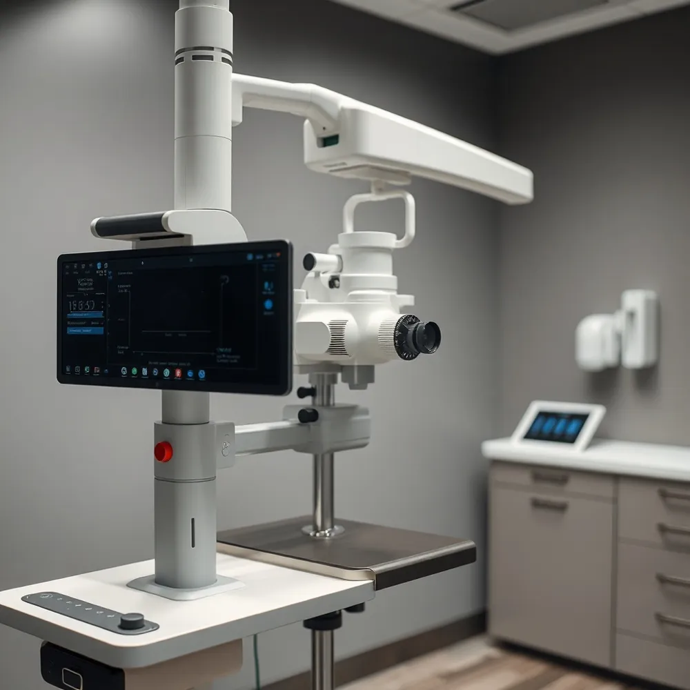
How modern diagnostics (DRI OCT Triton) aid early detection
Advanced imaging, such as the DRI OCT Triton, allows doctors to detect cataracts earlier than ever. This high-resolution optical coherence tomography reveals detailed layers of the lens, spotting subtle changes like posterior subcapsular plaques or early cortical spokes before they significantly impair vision. Early detection leads to better timing of treatment and improved outcomes. | Cataract Type | Key Feature | Typical Symptoms | |---|---|---| | Nuclear Sclerotic | Central lens hardening & yellowing | Blurred distance vision, myopic shift | | Cortical | Outer lens spoke-shaped opacities | Glare, halos, night vision problems | | Posterior Subcapsular | Back-of-lens plaque | Rapid vision loss, bright light glare |
Understanding the Spectrum of Cataract Types
Age-Related and Less Common Cataract Types
Age-related (senile) cataracts are the most common, with three subtypes: nuclear sclerotic (central lens hardening, yellowing, and myopic shift), cortical (peripheral wedge-shaped opacities causing glare), and posterior subcapsular (back-of-lens opacity disrupting reading and producing halos). Less common forms include traumatic (after eye injury), radiation (from UV or therapeutic irradiation), congenital (present at birth or in childhood), and secondary cataracts (posterior capsule opacification after surgery, treatable with laser). Risk factors that span all types include aging, diabetes, smoking, prolonged steroid use, UV exposure, and ocular trauma.
Nuclear Sclerotic Cataracts: The Most Prevalent Form
What is a cataract definition? Common cataract symptoms include glare and blurred vision. Three types of cataracts are nuclear sclerotic, cortical, and posterior subcapsular. Nuclear sclerotic cataract details involve lens hardening. Cortical cataract symptoms include spoke-like opacities. Posterior subcapsular cataract overview discusses risk factors. Age-related cataract types are common. Phacoemulsification cataract surgery is effective. Cataract surgery success rate is high. Real-world visual outcomes of cataract surgery show good results. WHO standards for postoperative visual acuity are used. Apple Eye Care cataract services provide care.
Cortical vs. Nuclear Cataracts: Key Differences
Anatomical Location and Appearance
Nuclear cataracts form in the central lens nucleus, causing gradual yellowing and hardening of the lens. Cortical cataracts develop in the outer cortex as white, wedge-shaped spokes that migrate inward toward the visual axis.
Visual Symptoms and Patient Complaints
Nuclear cataracts blur distance vision more than near, may temporarily improve reading (“second sight”), and reduce color discrimination. Cortical cataracts primarily produce glare—especially from headlights—along with blurred vision, halos, and diminished contrast and depth perception.
Risk Factors and Progression Patterns
Nuclear cataracts are strongly age-related and progress slowly over several years; smoking further increases risk. Cortical cataracts are more frequently linked to diabetes, ultraviolet exposure, and steroid use, and can progress more rapidly, with noticeable symptoms developing within months.
Posterior Subcapsular Cataracts: Distinct Features and Surgical Challenges
![]()
Location and Visual Impact
Posterior subcapsular cataracts (PSCs) form as an opacity on the posterior capsule, directly in the visual axis. Even small opacities cause pronounced glare, difficulty with near vision, and rapid visual decline.
Comparison with Cortical Cataracts
Unlike PSCs, cortical cataracts appear as peripheral wedge‑shaped opacities that initially spare central vision and progress slowly. PSCs are centrally located, more visually significant early, and strongly associated with steroid use, diabetes, and younger age.
Surgical Challenges
PSCs are surgically demanding because the dense plaque adheres to the posterior capsule, raising the risk of capsule rupture during phacoemulsification. Precise technique and careful planning are essential.
Risk of Blindness and Rapid Progression
Untreated PSCs can lead to blindness. They can progress quickly—structural changes have been observed within four weeks—necessitating timely surgical removal to restore vision and prevent severe impairment.
Serious Cataract Variants: Polar and Traumatic Forms
![]()
Which type of cataract is considered the most serious?
Posterior polar and traumatic cataracts are considered the most serious variants because of their higher surgical risk and complex visual impact. Posterior polar cataracts, often congenital, sit at the back of the lens and are linked to a fragile capsule, raising the chance of capsule rupture during surgery. Traumatic cataracts result from eye injury, may cause lens instability or dislocation, and require delicate, advanced surgical techniques. Management focuses on careful preoperative planning: for posterior polar, gentle techniques to avoid capsule stress; for traumatic, repairing associated damage and often delaying intraocular lens implantation. Early referral to an experienced surgeon is critical for optimal visual recovery.
Post‑Surgery Realities and the Latest Lens Innovations
![]()
What is the most frequent complaint after cataract surgery?
Increased light sensitivity and glare, especially in bright environments or with oncoming headlights, are the most common postoperative complaints. This occurs as the eye and new intraocular lens adjust during early healing.
How long do intra‑ocular lenses last?
Modern IOLs are made from durable, biocompatible materials and are designed to last a lifetime. They do not degrade. Some patients may develop posterior capsule opacification, a treatable clouding that is quickly resolved with YAG laser capsulotomy.
What are the newest cataract treatment options available in 2026?
Trifocal intra‑ocular lenses are the latest breakthrough, providing clear vision at near, intermediate, and far distances. This reduces dependence on glasses after surgery, representing a significant advance in personalized cataract care.
Putting It All Together for Clearer Vision
Recap of cataract type distinctions and why they matter
The three main cataract types—nuclear sclerotic, cortical, and posterior subcapsular—differ in where they form inside the lens and how they affect vision.
Nuclear sclerotic cataracts slowly yellow and harden the lens center, often temporarily improving near sight before blurring distance vision and dulling color perception. Cortical cataracts create spoke-like white opacities from the lens edges inward, typically causing disabling glare—especially from headlights at night. Posterior subcapsular cataracts develop rapidly at the back of the lens, directly in the visual path, making reading difficult and producing severe light sensitivity and halos.
Understanding which type you have helps your doctor predict how quickly vision may change and plan the best timing for treatment.
The importance of regular exams at Apple Eye Care
Because cataracts develop gradually, many people adjust to worsening vision without realizing it. Regular comprehensive eye exams at Apple Eye Care allow early detection of subtle lens changes.
Advanced diagnostics, such as the DRI OCT Triton, enable your doctor to assess the exact location and severity of lens opacities. This detailed picture helps distinguish cataract-related vision loss from other eye conditions, ensuring you receive the right advice at the right time.
How personalized IOL selection and advanced diagnostics improve outcomes
Cataract surgery replaces the cloudy lens with a clear intraocular lens (IOL). Modern IOL choices include monofocal, multifocal, toric, or accommodative designs.
Your surgeon at Apple Eye Care uses precise pre-operative measurements—axial length, corneal power, and anterior chamber depth—to select an IOL tailored to your visual needs and lifestyle. This personalized approach helps reduce dependence on glasses and optimizes vision at all distances.
Advanced surgical techniques, such as laser-assisted cataract surgery, further improve accuracy and recovery. With these tools, Apple Eye Care aims to deliver the excellent outcomes reported nationally, where over 97% of procedures restore clear vision.
| Cataract Type | Location in Lens | Key Visual Symptom | Typical Progression |
|---|---|---|---|
| Nuclear sclerotic | Centre (nucleus) | Dulling of colour, blurred distance | Slow (years) |
| Cortical | Outer edge (cortex) | Glare, especially at night | Variable (months to years) |
| Posterior subcapsular | Back surface (under capsule) | Reading difficulty, light sensitivity | Rapid (months) |