Seeing Beyond the Standard Exam
For many patients, the first question about an OCT (Optical Coherence Tomography) scan is straightforward: what does it cost, and is it worth it? The answer depends on how you value seeing a specialist who catches problems early. At Apple Eye Care, we believe in transparency about both the clinical value and the out-of-pocket expense, and we design our pricing to keep advanced diagnostics accessible to every family in our practice.
Let’s break down what you’re paying for. A standard eye exam checks visual acuity, refraction, and basic eye health. An OCT scan goes further: it uses light waves to create a cross-sectional image of the retina, optic nerve, and macula. This is the same technology found in the Triton OCT used in advanced diagnostic centers, and it’s what allows us to see the layers of your eye that a traditional exam simply cannot reveal.
Optical Coherence Tomography Unlocks the Retina
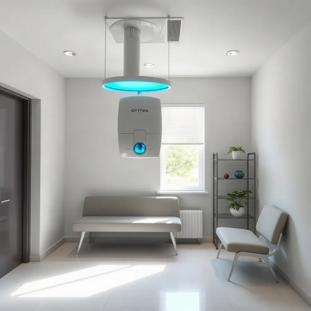 Optical coherence tomography (OCT) is a powerful diagnostic tool, but it is not without practical limitations. The most significant constraint is that standard OCT devices, like the DRI OCT Triton, are designed to capture high-resolution cross-sectional images of the retina, which requires the patient to remain still and cooperate with the scan. This can be challenging for young children, individuals with involuntary eye movements (nystagmus), or patients who have difficulty positioning their chin and forehead against the device's rest. In such cases, image quality may be compromised, reducing the reliability of the scan.
The Challenge of Media Opacities and Motion Artifacts
Even with advanced technology, OCT has limitations when ocular media are not clear. Dense cataracts, severe corneal scarring, or vitreous hemorrhage can scatter light before it reaches the retina, degrading image resolution. This means that in some patients, the very condition that requires advanced diagnostic imaging may obscure the very structures the test aims to visualize. Additionally, patient movement during image acquisition, such as a heartbeat, breathing, or a simple blink, can introduce motion artifacts that create false or distorted areas in the cross-sectional scans. While newer devices employ eye-tracking and image-averaging algorithms to mitigate this, motion artifact remains a real-world concern for all optical coherence tomography platforms.
Interpretation Requires Specialized Expertise
Another limitation is that OCT does not provide a diagnosis by itself. The automated retinal nerve fiber layer (RNFL) and ganglion cell analysis (GCA) reports generate, but they require a clinician to interpret them in the context of the patient's visual field test and clinical history. A single suspicious surface-area map can be a normal variant, a sign of glaucoma, or an artifact. Therefore, the accuracy of the test heavily depends on the interpreting doctor's training. At a family-focused practice like Apple Eye Care, the advanced diagnostic technology is only as effective as the clinical team that analyzes the scans.
Cost and Accessibility
Cost and accessibility are also notable limitations. Advanced OCT units, such as the Topcon DRI Triton with its swept-source technology, represent a substantial capital investment for an eye care practice. For patients, this sometimes translates into a higher out-of-pocket cost for an advanced diagnostic scan, especially if insurance providers consider it non-covered or a non-basic service. Furthermore, not every clinic has the volume or the specialist expertise to justify or operate such machinery. This is precisely why Apple Eye Care has integrated these technologies into its examination suite, offering patients access to advanced diagnostics that many general practices lack, within a setting that does not require referral to a hospital.
| Limitation | Detail | Impact on Patient Care |
|---|---|---|
| Motion Artifacts | Eye movement can distort images | Repeat scans required/less reliable data |
| Media Opacities | Scarring or cataracts block light | Reduces image resolution/quality |
| Interpretation Expertise | Needs specialized clinician analysis | Clinical outcome depends on the reader |
| Cost and Access | High-tech equipment is expensive | May not be available at every practice |
Why These Limitations Matter for Your Eye Health
Understanding these limitations does not diminish the value of OCT. Rather, it highlights the importance of choosing an eye care specialist who has the expertise to work around them. At Apple Eye Care, the clinical team is trained to recognize when artifacts or poor-quality scans compromise the data and will not hesitate to repeat the test, use alternative diagnostic tools, or correlate findings with your visual field. This personalized attention ensures that a limitation in technology never becomes a limitation in your care.
Wide-Field Retinal Imaging and Visual Field Testing
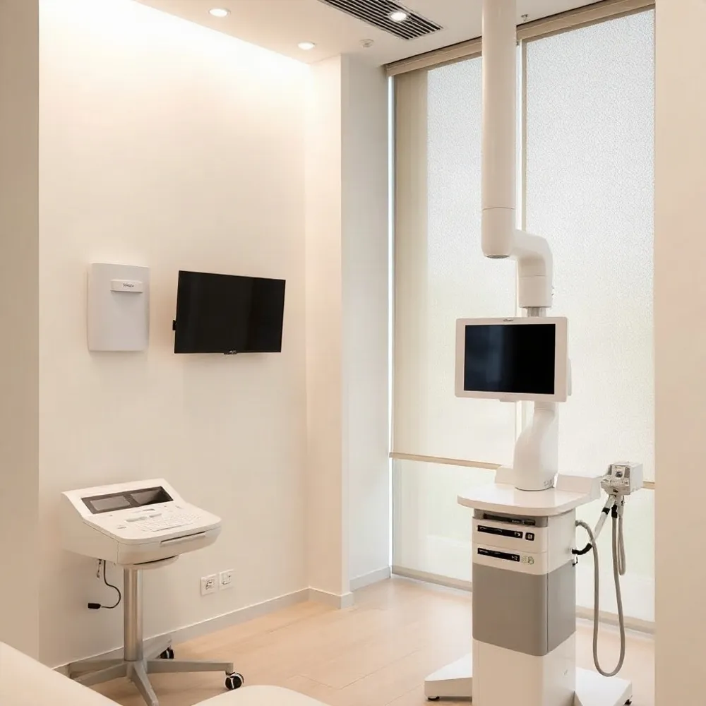 Wide-field retinal imaging has become a standard diagnostic tool in modern eye care, but its real value at Apple Eye Care shows up in how it changes the everyday exam. A single ultra-widefield image captures more than 80% of the retina in one capture, compared with the roughly 15% visible through a traditional direct ophthalmoscope. For patients, that means a more thorough baseline without the need for dilating drops in many cases. For the doctor, it means seeing peripheral lesions, lattice degeneration, and diabetic changes that would otherwise go unnoticed until they cause symptoms.
The clinical decisions that follow a wide-field image often hinge on volume and precision. Optos popularized the ultra-widefield category with its scanning laser ophthalmoscope, which produces a single panoramic view of the retina. But the category has since expanded: Topcon's DRI OCT Triton combines swept-source OCT with angiography to show both structure and blood flow in the same device. At Apple Eye Care, the DRI OCT Triton is the workhorse for advanced diagnostics, offering 1050 nm wavelength imaging that penetrates deeper into the choroid than older spectral-domain systems. That depth matters for glaucoma staging and for spotting subretinal fluid that other devices miss.
Where the platforms separate is in integration and workflow. Optos excels at a single, fast capture that triages well in a high-volume practice. Topcon's strength is the multimodal fusion of OCT, OCT-A, and fundus photography in one unit. Apple Eye Care pairs the wide-field screening capability with the Triton's detailed cross-sectional scans, so a patient who needs a closer look at the optic nerve or macula can be assessed without leaving the exam lane. The result is a diagnostic pathway that starts with a broad retinal photo and narrows to specific layers, all in under 10 minutes.
Scope. Optos captures wide-field images (up to 200 degrees) to screen the peripheral retina; the Triton's OCT sections through the central retina and optic nerve for layer-by-layer analysis.
Integration. Optos offers a standalone retinal camera; Topcon's multimodal platform combines OCT, OCT-A, and fundus photography. Apple Eye Care uses the Triton's all-in-one approach to reduce patient time.
Intelligence. Artificial intelligence algorithms on some platforms flag suspected glaucoma or macular pathology automatically. At Apple Eye Care, the doctor reviews the Triton's high-density scans to confirm findings before treatment.
The practical difference shows up in patient experience. With an Optos-style device, most patients avoid dilation drops and get an immediate full-color image. But that image is a photograph; it does not show the internal layers of the retina. The Triton's OCT adds that second dimension, which is why Apple Eye Care combines both technologies. A patient with diabetes who needs annual screening gets the wide-field view for peripheral ischemia, then a quick OCT to check for macular edema. Both are non-invasive, both are covered by most insurances, and both are performed in the same visit.
Choosing between these systems depends on the practice's focus. A high-volume primary care clinic might standardize on Optos for speed. A specialty practice that manages glaucoma and macular disease needs the OCT's quantitative data. Apple Eye Care falls into the second category. The practice's investment in the Triton reflects its advanced diagnostic testing philosophy: catch problems early, monitor progression with precise scans, and treat before vision loss occurs.
| Feature | Optos | Topcon DRI OCT Triton | Apple Eye Care approach |
|---|---|---|---|
| Capture type | Ultra-widefield fundus photo | Swept-source OCT + OCT-A | Both, as needed |
| Retinal coverage | Up to 200 degrees | Posterior pole + wide-field OCT | Single-visit full workup |
| Dilation | Often not needed | Not needed | Often not needed |
| Best for | Screening, peripheral lesions | Glaucoma, macular pathology | Diagnosis and monitoring |
Corneal Mapping and Dry Eye Diagnostics
Optical coherence tomography (OCT) is one of the most significant advances in ophthalmic imaging. Per a 2023 review in The Lancet Global Health, AI‑enhanced OCT is reshaping how clinicians detect and manage retinal and optic nerve disease. This non‑invasive, high‑resolution imaging is a core diagnostic tool for conditions that often go unnoticed in a standard eye exam.
Scope. OCT captures cross‑sectional images of the retina, optic nerve, and cornea. While a standard exam documents visual acuity and anterior segment health, OCT adds micrometer‑level detail of tissue layers, revealing early signs of glaucoma, diabetic macular edema, and age‑related macular degeneration.
Integration. Modern OCT systems pair with fundus photography and visual field testing to create a composite view. This integration lets a clinician correlate structural changes with functional vision loss, producing a far more complete assessment than any single test alone.
Intelligence. Advanced platforms such as the DRI OCT Triton and Topcon's OCT‑A incorporate swept‑source technology for faster acquisition and deeper penetration. On the AI front, a 2023 systematic review found that deep learning models can accurately segment retinal layers and identify pathology, supporting earlier intervention and reducing variability in diagnosis.
AI, Future Tech, and the Human Eye Care Team
Artificial intelligence is quietly changing how eye doctors detect disease. A 2023 Lancet review noted that AI and digital health tools are increasingly used in global eye health, from screening to diagnosis to monitoring chronic conditions. Artificial intelligence and digital health in global eye health shows how algorithms now help interpret retinal scans with remarkable consistency, flagging early signs of diabetic retinopathy, glaucoma, and age-related macular degeneration before they cause noticeable vision loss.
At Apple Eye Care in El Paso, TX, that technology is not a future concept. The practice uses advanced diagnostic technology such as the DRI OCT Triton, a swept-source OCT that captures ultra-detailed cross-sections of the retina and optic nerve. This allows Dr. Stephen Applebaum and his team to spot subtle structural changes months or years earlier than traditional methods, often before any symptoms appear.
What Makes New Diagnostic Devices Different
Standard eye exams rely on subjective tests: reading letters, pushing a button when you see a flash. Modern diagnostic devices add objective, quantitative data. Optical coherence tomography (OCT) measures retinal thickness in microns. Visual field analyzers map your peripheral vision precisely. Corneal topographers build a 3D map of your eye's front surface. Together, these tools detect conditions that a routine refraction simply cannot see.
Apple Eye Care incorporates AI-smart technology in eye exams to automate and enhance this analysis. Instead of relying solely on a doctor's visual inspection of images, AI algorithms compare scans against large datasets of normal and diseased eyes. They flag suspicious regions for closer review, reducing the chance of human oversight. The result is a more consistent, data-backed assessment that supports, rather than replaces, clinical judgment.
Comparing Diagnostic Capabilities
| Technology | What It Measures | Typical Use |
|---|---|---|
| OCT (Optical Coherence Tomography) | Retinal and optic nerve structure in microns | Detecting glaucoma, macular edema, AMD |
| Visual Field Analyzer | Peripheral sensitivity thresholds | Monitoring glaucoma progression |
| Corneal Topography | Anterior corneal curvature | Planning LASIK, fitting hard contact lenses |
| Fundus Photography | High-res images of the retina | Documenting diabetic retinopathy, drusen |
The DRI OCT Triton series from Topcon, for example, uses swept-source technology to image deeper into the eye than traditional spectral-domain OCT. This can reveal choroidal detail that matters in conditions like central serous retinopathy or age-related macular degeneration. Apple Eye Care chose this device for its ability to provide precise, reproducible measurements, which directly informs treatment decisions for cataract and glaucoma management.
Why Early Detection Matters
The most insidious eye diseases are silent until they are advanced. Glaucoma can steal peripheral vision slowly, and by the time you notice, up to 40% of optic nerve fibers may already be lost. Diabetic retinopathy often shows no symptoms until the retina bleeds. Catching these changes early is the single best way to preserve vision. The role of technology in eye exams and vision screenings explains how modern tools now allow this earlier detection reliably.
Apple Eye Care integrates these diagnostics into every comprehensive exam. When a scan reveals a suspicious lesion or unexpected thinning, the team can act fast, whether that means scheduling a same-day follow-up, referring to a specialist, or beginning treatment in-house. This proactive approach contrasts with clinics that reserve advanced imaging for already-symptomatic patients, where treatment options are more limited.
Combining AI with Human Judgment
AI is a powerful assistant, but it is not a replacement for an experienced eye doctor. The algorithms are trained on millions of images, but they can miss rare conditions or misinterpret artifacts. Dr. Applebaum and his team combine the quantitative outputs of AI with their clinical experience and the patient's individual history. The goal is not to outsource decision-making, but to enhance it. This balanced approach means you get a more thorough assessment, without losing a human touch.
For patients, the benefit is tangible: a shorter wait for answers, more precise monitoring of chronic conditions like glaucoma, and greater confidence that nothing has been missed. Apple Eye Care makes this technology accessible without extra cost to standard exam patients, which sets it apart from clinics that charge separately for every advanced scan.
The Future of Vision Care
Looking ahead, the trend is toward even more integrated and accessible care. From portable OCT devices that can be used outside the clinic to AI that can predict progression risk, optometry is moving earlier. The future of vision and eye care points toward a model where these tools are standard, not exceptional. Apple Eye Care is positioned to stay ahead of that curve, continuously updating its equipment and training so that El Paso families receive care that feels personal and precise.
Your Eyes Deserve Advanced Screening
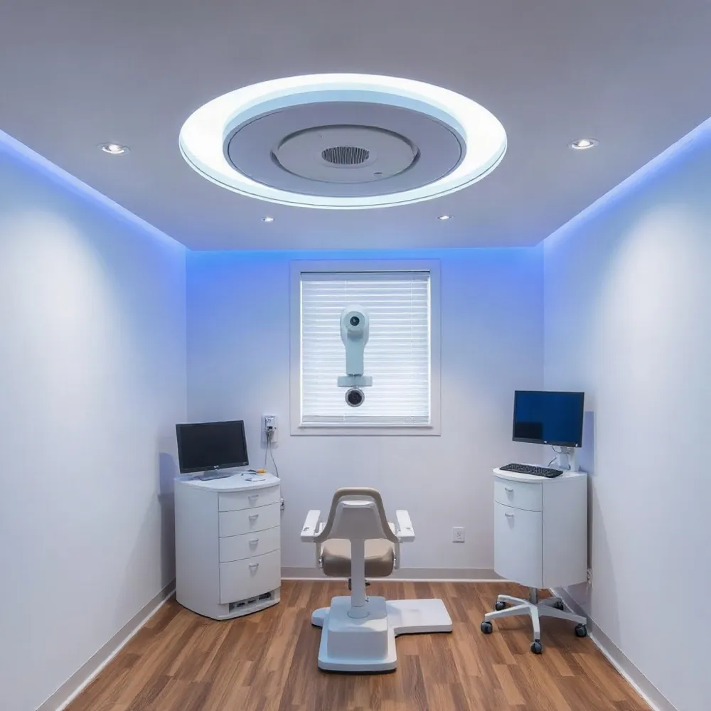 Modern optometry has moved well beyond the basic eye chart. Advanced diagnostic technology now lets eye care professionals inspect the structures of your eyes in ways that were once reserved for specialists. Devices like the DRI OCT Triton from Topcon use swept-source optical coherence tomography (OCT) to capture detailed cross-sectional images in seconds, helping detect conditions such as glaucoma, macular degeneration, and diabetic retinopathy at their earliest, most treatable stages. Sources like the The Lancet's review of AI in digital eye health confirm that these technologies are reshaping how eye diseases are found and managed worldwide.
For patients, the difference is tangible. Instead of relying only on the familiar 'which is better, one or two' routine, you can now undergo scans that map your retina and optic nerve with precision. The advanced diagnostic tests offered through your optometrist can reveal signs of disease before you notice any symptoms. This is especially important because common vision threats like glaucoma and age-related macular degeneration often develop silently, with no pain or warning signs until significant vision has already been lost.
What Counts as Advanced Diagnostic Technology?
Several technologies fall under the advanced diagnostic umbrella, each designed to give a clearer picture of your ocular health:
- Optical coherence tomography (OCT) creates cross-sectional images of the retina and optic nerve, allowing for precise measurement of tissue thickness and early detection of conditions like glaucoma and macular edema.
- Automated visual field testing maps your peripheral vision to detect blind spots that may signal nerve damage.
- Digital retinal photography and fundus imaging capture detailed records of your retina's appearance, enabling side-by-side comparison over time.
- Wavefront aberrometry measures how light travels through your eye, aiding in precise vision correction and contact lens fitting.
Practices that invest in such equipment, including Apple Eye Care in El Paso, TX, use these tools to provide a level of insight that a standard exam alone cannot. When you visit a practice equipped with modern imaging, you benefit from earlier detection and more tailored care.
How Apple Eye Care Uses This Technology
At Apple Eye Care, advanced diagnostics are part of the standard approach to patient care. The practice uses the DRI OCT Triton from Topcon, which provides swept-source OCT imaging and can also capture anterior segment details used in cataract and glaucoma management. This means that during a routine visit, your optometrist can assess not only your prescription but also the health of the tissue layers that matter most. The detailed scans help guide decisions about dry eye treatment, monitoring of cataracts, and management of glaucoma, all within a family-friendly setting.
The practical value is clear: you leave the exam with more than just a prescription. You get a conversation about your eye health, supported by images and measurements your provider can explain. For busy families in El Paso, that efficiency translates to less time in the chair and more confidence in the care you receive.
Comparing Advanced Diagnostics: Key Technologies at a Glance
To understand what sets modern equipment apart, it helps to see the differences side by side:
| Technology | What It Does | What It Helps Detect |
|---|---|---|
| OCT (Swept-Source) | Cross-sectional imaging of retina and optic nerve | Glaucoma, macular degeneration, diabetic retinopathy |
| Visual Field Testing | Maps peripheral vision | Glaucoma, neurological issues |
| Digital Retinal Photography | High-res photo of the back of the eye | Retinal tears, diabetic changes |
| Wavefront Aberrometry | Measures how light focuses in the eye | Refractive error, cataract planning |
Why This Matters for Your Eye Exam
The technology itself is only half the story. What matters is how your eye care team uses it. A practice that integrates advanced diagnostics into routine care gives you a significant advantage: problems can be spotted earlier, treatment can be more precise, and you can be more confident in the advice you receive. That is exactly the philosophy at Apple Eye Care, where the combination of modern equipment and a compassionate, family-focused team means you are not just another patient in the chair.
A Clearer Path Forward
When it comes to staying current with eye care technology, a 2020 review in The Lancet highlighted how digital health tools, including OCT, are transforming global eye health. Artificial intelligence and digital health in global eye health describes how these advances enable earlier detection and more precise treatment planning. At Apple Eye Care in El Paso, we integrate this kind of innovation to bring you care that goes beyond the standard exam, and we're proud to offer advanced diagnostics that help protect your vision for years to come.
Advanced diagnostic technology like OCT has become a crucial part of modern optometry. According to the Advanced Diagnostics guide, these tools allow optometrists to see layers of the retina in detail that wasn't possible a generation ago. This deeper look means we can catch conditions like glaucoma, macular degeneration, and diabetic retinopathy earlier, often before you notice any symptoms. By incorporating such technology into routine eye exams, Apple Eye Care families in El Paso benefit from personalized care that prioritizes early detection.
What Advanced Diagnostic Technology Means for You
Advanced diagnostic technology typically includes tools like Optical Coherence Tomography (OCT) and fundus photography. These machines take high-resolution cross-sectional images of your retina, optic nerve, and blood vessels, giving your eye doctor a clear picture of your eye health. At Apple Eye Care, we use this technology to assess the thickness of your retinal nerve fiber layer and identify subtle changes that might indicate early-stage glaucoma. This proactive approach means we can begin treatment sooner, potentially preserving more of your vision.
For families, this technology offers peace of mind. Whether it's checking for the signs of diabetic eye disease or monitoring a family history of macular degeneration, advanced diagnostics give us the data we need to make informed decisions about your eye health. At Apple Eye Care, we're committed to making these sophisticated exams accessible and comfortable, so you can feel confident that every aspect of your vision is being thoroughly examined.
The Role of AI in Modern Eye Exams
Artificial intelligence is another innovation shaping the future of eye care. AI smart technology in eye exams can analyze OCT scans with remarkable speed and accuracy, flagging potential issues that might be missed by the human eye. This doesn't replace your optometrist; rather, it acts as an extra set of eyes, ensuring nothing is overlooked. At Apple Eye Care, we're excited about how these tools can support early intervention and improve outcomes for our patients in El Paso.
Advanced Imaging Tools We Use
One of the leading technologies we use is the DRI OCT Triton from Topcon Healthcare. This multimodal swept-source OCT provides detailed images of the retina and choroid, the layer beneath the retina, which is essential for detecting conditions like wet age-related macular degeneration. Its high-speed imaging captures data in seconds, making the exam comfortable and efficient for you and your family.
Another tool in our advanced diagnostic suite is the Optos ultra-widefield imaging system. This allows us to capture a 200-degree view of your retina in a single image, revealing peripheral vision problems that traditional exams might miss. Combined with our advanced diagnostic technology, these tools provide a comprehensive evaluation of your eye health.
| Technology | What It Does | Conditions Detected |
|---|---|---|
| OCT (DRI OCT Triton) | High-res cross-sectional retina images | Glaucoma, AMD, diabetic retinopathy |
| Ultra-widefield imaging | Full retina view in one shot | Peripheral retina tears, detachment |
| AI analysis | Flags potential issues in scans | Early signs of disease progression |
Empowerment Through Early Detection
When you understand your eye health, you can take proactive steps to maintain it. Advanced diagnostics empower you with the knowledge that comes from early detection. For instance, if we detect elevated intraocular pressure or a thinning optic nerve, we can intervene with treatments that slow the progression of glaucoma, potentially saving you from significant vision loss. At Apple Eye Care, we're dedicated to giving you the personalized attention that turns complex data into a clear care plan.
We also recognize that staying current with technology is essential in today's fast-paced world. That's why we invest in the latest diagnostic systems and train our team accordingly. When you choose Apple Eye Care, you're choosing a practice that puts your vision first, backed by tools that are at the forefront of optometric medicine.