The Evolution of Precision in Eye Care
The field of optometry is undergoing a significant transition from reactive assessment to proactive, data-driven management. Traditional diagnostic methods often provide limited snapshots of eye health, whereas modern standards rely on high-resolution, multimodal imaging to identify subtle structural changes Nature early.
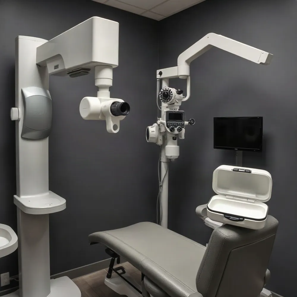
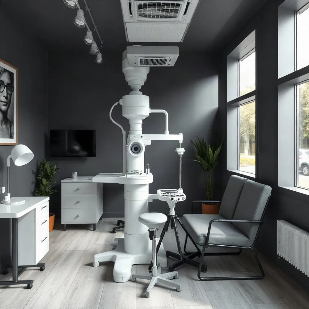
At Apple Eye Care in El Paso, TX, the commitment to this precision is reflected in the integration of the DRI OCT Triton. By utilizing swept-source optical coherence tomography, this device achieves deep penetration through ocular media such as cataracts, capturing detailed visualizations of both the macula and optic nerve in seconds Retinal Physician. This level of detail allows for more accurate longitudinal tracking of conditions like glaucoma Glaucoma Foundation.
This technological shift ensures that patients receive care tailored to their unique ocular anatomy. By combining advanced diagnostics with consistent patient monitoring, practices can now address potential issues before they cause permanent vision loss Stanford Medicine.
Diagnostic Innovations at Apple Eye Care
- The DRI OCT Triton utilizes 1,050 nm swept-source technology to image through cataracts.
- The device achieves a capture speed of 100,000 A-scans per second to minimize motion artifacts.
- Widefield imaging captures macula and optic nerve data in one 1.8-second scan.
- Artificial intelligence facilitates early disease detection and prioritized patient triage.
- The iCare HOME2 enables longitudinal tracking of intraocular pressure outside clinical settings.
- OCT Angiography maps retinal vasculature without requiring invasive dye injections.
- Polygenic risk scoring identifies patient genetic susceptibility to glaucoma before symptom onset.
- Femtosecond lasers replace manual incision methods to automate and refine cataract surgery.
- Intraoperative aberrometry provides real-time refractive analysis to optimize lens selection.
- Adaptive optics research offers potential for cellular-level visualization of retinal structures.
1. Swept Source Optical Coherence Tomography Imaging
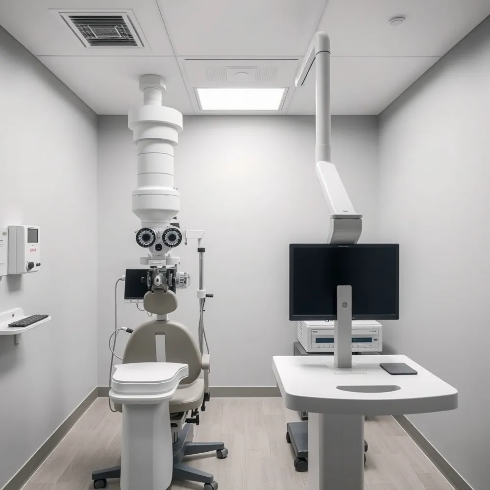 Modern eye diagnostics rely on high-resolution imaging to detect subtle pathologies before they manifest as vision loss. At Apple Eye Care, the DRI OCT Triton utilizes swept-source technology to provide a foundational diagnostic advantage. Unlike traditional spectral-domain systems, this device employs a 1,050 nm wavelength that offers significantly deeper tissue penetration.
This specialized light source is particularly effective at capturing clear images through media opacities, such as cataracts or vitreous hemorrhages, which often obscure view in standard exams. By operating at a speed of 100,000 A-scans per second, the technology reduces the impact of involuntary ocular movement and microsaccades. This improved efficiency minimizes the need for repeated scans, ensuring that practitioners obtain reproducible data for accurate longitudinal monitoring of glaucoma and retinal health.
The use of invisible scanning light also enhances patient experience by eliminating the distraction of visible light lines during the capture process. This combination of speed, deep penetration, and superior image quality allows for more confident clinical decision-making when managing complex cases in El Paso.
2. Wide Field Diagnostic Scan Capabilities
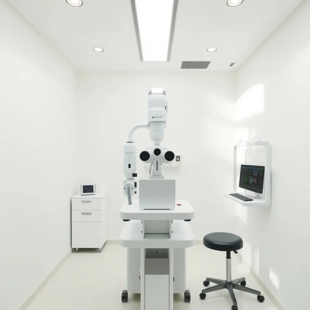 Traditional eye diagnostics often required separate, time-consuming scans for the macula and optic nerve. By using Widefield OCT imaging, clinicians can now capture both regions in a single 12 mm x 9 mm scan. At Apple Eye Care, the use of the DRI OCT Triton exemplifies this efficiency, as it simultaneously records high-resolution data for both areas in just 1.8 seconds.
This synchronized approach provides significant benefits for patient comfort. Because patients spend less time in the chair and do not need to repeat imaging, the impact of motion, alignment errors, and fatigue is substantially minimized per a 2023 study in Nature. Furthermore, wide-field scans are superior in capturing clinically relevant defects, including those in the peripheral regions at the 11 and 12 o'clock positions that smaller cube scans might miss.
The DRI OCT Triton stands out from older spectral-domain systems by using a 1,050nm wavelength to penetrate media opacities more effectively. This ensures that even patients with early-stage cataracts receive accurate, reproducible measurements, allowing for reliable monitoring of retinal ganglion cell loss over time.
3. Artificial Intelligence in Retinal Screening
 Artificial Intelligence is shifting glaucoma management from reactive assessment to a proactive model by applying deep learning to multimodal datasets. At Apple Eye Care, we use the DRI OCT Triton to generate high-resolution structural data that feeds into these automated systems, ensuring that our patients benefit from the latest innovations in diagnostic accuracy.
Deep learning models process diagnostic data similarly to the human brain, offering faster and more precise detection of glaucomatous patterns in ways that traditional clinical tools like the standard slit lamp cannot match. By analyzing these complex datasets, AI can help clinicians determine the optimal timing for intervention or surgery through individualized risk assessments, a task often difficult to achieve with static snapshots taken during office hours.
Scalability and triage efficiency are primary drivers for adopting these models in busy clinical environments. Automated systems like RETFound demonstrate the ability to detect disease before clinical symptoms even manifest. This allows practices to prioritize high-risk patients efficiently, ensuring that limited resources are directed where they provide the most substantial impact on preserving vision.
4. Remote Home Tonometry Systems
Traditional clinical practice relies on office-based measurements, which provide only a single snapshot of intraocular pressure that may miss significant fluctuations occurring throughout the day. Because pressure often reaches its peak during early waking hours, many patients remain at risk despite seemingly normal values during standard examinations per New Technology Offers Modern Canary in the Glaucoma Coal Mine.
The iCare HOME2 system addresses these variations by allowing individuals to monitor their pressure independently outside of professional settings. By capturing time-stamped, longitudinal data, this tool helps clinicians identify treatment failures, such as ineffective medication or inadequate surgery, far faster than traditional biannual visits. At Apple Eye Care, we integrate advanced diagnostics like the DRI OCT Triton to monitor structural changes, which complements the functional insights gained from home tonometry in identifying shifts like early morning spikes.
Continuous remote monitoring fundamentally shifts the patient role from passive recipient to active participant in their own care. This process empowers individuals to understand how daily activities, lifestyle factors, and medication adherence directly impact their ocular health. As noted by EYEWORLD, this transition toward proactive management is vital for preventing disease progression.
5. Optical Coherence Tomography Angiography
Optical Coherence Tomography Angiography provides a non-invasive, dye-free method to visualize blood flow within the eye. By detecting the movement of red blood cells, this technology maps the retinal and choroidal vasculature without requiring the traditional, invasive injections used in conventional angiography.
For clinicians at Apple Eye Care, utilizing advanced diagnostic tools is essential for early detection. Unlike standard imaging, which focuses on structural thickness, this method is especially valuable for tracking disease progression in severe glaucoma. Because vessel density measurements are capable of remaining stable longer than traditional structural markers, they provide a clearer picture when a patient's results reach a clinical floor.
Integrated diagnostic platforms, such as the DRI OCT Triton, allow for the seamless incorporation of these vascular maps into routine practice. This level of precision helps eye care providers distinguish between healthy physiological variations and early signs of pathology, ensuring more accurate treatment plans for complex ocular conditions.
6. Genetic Polygenic Risk Scoring
The evolution of glaucoma care now allows clinicians to assess a patient's inherited susceptibility before visible symptoms arise. Polygenic risk scoring analyzes multifaceted genetic data to identify individuals with a high predisposition to disease, particularly when a family history is present but clinical imaging currently appears normal. By determining these risk profiles early, eye care professionals can transition from standard screening schedules to a personalized monitoring strategy that prioritizes high-risk patients for more frequent observation.
At Apple Eye Care, we focus on combining state-of-the-art diagnostic tools like our DRI OCT Triton with emerging genetic insights. While tools like polygenic risk scores help quantify an individual's vulnerability to condition development, proactive intervention remains essential. Unlike traditional reactive models that only respond to damage, these genetic indicators allow for targeted follow-up intervals, ensuring that early signs of glaucoma progression are caught during the earliest, most manageable stages.
7. Femtosecond Laser Assisted Cataract Surgery
Modern cataract intervention has moved beyond traditional manual techniques toward highly precise laser platforms. Femtosecond laser assisted cataract surgery uses ultra-short laser pulses to automate critical surgical steps, including corneal incisions and lens fragmentation. By replacing manual blade incisions with laser guidance, surgeons minimize human error and achieve higher levels of accuracy during the procedure.
At Apple Eye Care in El Paso, TX, delivering predictable visual outcomes is a priority. While some centers rely solely on conventional phacoemulsification, our practice integrates advanced pre-surgical diagnostics like the DRI OCT Triton to map individual ocular anatomy. This data-driven foundation ensures that laser-assisted techniques are tailored to the specific structure of the patient eye.
This technology is especially beneficial for patients seeking premium intraocular lens (IOL) implantation. Precise incisions and centered lens positioning are essential for complex lenses that correct astigmatism or presbyopia. The combination of consistent laser-guided automation and personalized diagnostic imaging helps reduce complications, while supporting faster recovery times for the patient.
8. Intraoperative Aberrometry for Lens Selection
Selecting the optimal intraocular lens (IOL) during cataract surgery is a precise process that directly impacts a patient's post-operative visual clarity. Apple Eye Care supports this level of procedural precision by integrating advanced diagnostic data throughout every stage of eye health management. Unlike traditional preoperative calculations that may rely on static biometric measurements, intraoperative aberrometry provides real-time refractive power analysis while the patient is still on the surgical table.
Systems like the ORA (Optiwave Refractive Analysis) system utilize this wavefront technology to measure the eye's refractive state after the clouded lens has been removed. By capturing data at this specific moment, surgeons can refine the IOL power selection and verify axis alignment for toric lenses immediately. This approach significantly enhances the accuracy of IOL selection and positioning, which is vital for achieving the specific visual goals of patients seeking minimized dependence on glasses after their procedure.
Personalized outcomes for cataract patients are further optimized by combining these real-time surgical measurements with detailed preoperative diagnostics like the DRI OCT Triton. While competitors may depend on estimations based strictly on pre-procedure exams, this combination of diagnostic imaging and intraoperative aberrometry allows for a highly customized treatment path that accounts for the specific anatomy and unique refractive characteristics of each eye.
9. Adaptive Optics High Resolution Imaging
The pursuit of high-resolution diagnostics reaches its current architectural limit through Adaptive Optics scanning systems. By employing deformable mirrors to correct for ocular aberrations, this technology provides cellular-level visualization of retinal layers that standard clinical tools often fail to resolve. While traditional methods offer a broad overview of health, adaptive optics allows clinicians to view the posterior segment structures, such as the retinal nerve fiber layer bundles or the lamina cribrosa, with unprecedented structural detail.
This precision is vital for identifying microvascular changes early on, often before conventional imaging can detect subtle indications of disease. Current standard imaging, such as the DRI OCT Triton used at Apple Eye Care, remains the foundational choice for routine cataract and glaucoma management in El Paso due to its balance of reliability and deep-tissue penetration. In contrast, researchers are exploring the experimental potential for microscopic diagnostics within adaptive optics to refine how we track disease onset. While high barrier-to-entry costs currently restrict these systems primarily to research settings, they pave the way for a future where clinicians could monitor individual neuron health as a routine component of preventative eye care.
Foundational Diagnostic Tools for Eye Health
Providing specialized care for conditions like cataracts and glaucoma requires an integrated approach to early detection and routine monitoring. At Apple Eye Care in El Paso, TX, the DRI OCT Triton serves as a foundational diagnostic resource. This platform utilizes 1,050-nm swept-source technology to bypass media opacities, allowing for detailed imaging of the retina and optic nerve that standard devices might miss.
What tools and diagnostics are used to diagnose cataracts and glaucoma?
Cataract diagnosis begins with a comprehensive slit lamp biomicroscope exam to inspect the lens for clouding. Glaucoma management involves a more rigorous diagnostic process, starting with tonometry to measure intraocular pressure. Clinicians also employ pachymetry for corneal thickness and gonioscopy to examine the drainage angle of the eye. To track structural nerve fiber layer thinning, practitioners use Optical Coherence Tomography to generate high-resolution diagnostic maps.
Functional assessment remains equally vital to this standard workflow. Patients undergo automated perimetry to identify visual field deficits caused by optic nerve damage. By combining these structural and functional insights, providers can establish a clearer baseline for longitudinal care, helping to distinguish between stable ocular health and active disease progression.
Integrating OCT into Patient Care
Optical Coherence Tomography (OCT) is a vital non-invasive imaging tool that uses light waves to capture detailed, three-dimensional, cross-sectional images of the retina and the optic nerve. By allowing eye care professionals to map and measure the thickness of these distinctive layers, OCT enables the early detection and proactive management of serious conditions like glaucoma, macular degeneration, and diabetic retinopathy.
What is the role of OCT technology in comprehensive eye exams?
Because many of these diseases can develop without noticeable symptoms, regular OCT scans are essential for identifying changes in eye health over time. Furthermore, the technology provides clear, visual representations that help patients better understand their own eye health and treatment needs. At Apple Eye Care, incorporating advanced diagnostics ensures we provide the highest standard of personalized, preventative vision care. Unlike older spectral-domain systems, the Triton utilizes a 1,050nm wavelength, allowing our team to penetrate through mature cataracts to obtain high-resolution diagnostic data that might otherwise be obscured.
Future Shifts in Cataract and Glaucoma Therapies
Cataract surgery continues to evolve through improved intraocular lens technology, offering premium options that correct presbyopia and astigmatism for better visual outcomes. At Apple Eye Care, we integrate these advancements by using precise diagnostic data from the DRI OCT Triton to ensure that every lens selection and treatment plan is tailored to the specific ocular anatomy of our patients.
In glaucoma care, a shift toward interventional management is growing, with surgeons now using Minimally Invasive Glaucoma Surgeries and advanced laser therapies to optimize pressure control. Pharmaceutical developments are also expanding, with new long-acting drug delivery systems and once-daily formulations aimed at improving patient adherence.
Looking ahead, research into neuroprotective therapies is gaining momentum, potentially offering ways to preserve vision beyond simple pressure reduction. Furthermore, AI-driven diagnostic tools are emerging to better detect disease progression by analyzing imaging and clinical data. These innovations reflect a significant transition in ophthalmology, where Apple Eye Care utilizes modern technology to provide proactive management rather than relying on traditional diagnostic methods alone.
Commitment to Advanced Eye Care Success
Modern diagnostic tools and proactive management have fundamentally reshaped eye health by shifting the focus from crisis response to long-term preservation. Technology such as the DRI OCT Triton allows clinicians to detect subtle anatomical changes before they compromise sight, providing a clear pathway for better longitudinal health outcomes.
At Apple Eye Care in El Paso, we prioritize these innovations to ensure our patients receive the highest standard of precision. By integrating high-resolution Swept-Source OCT and personalized screening protocols, we create tailored management plans that safeguard vision for years to come. Patients are encouraged to visit www.appleeyecare.com to schedule an examination and experience how our commitment to modern, data-driven eye care supports your specific ocular needs.