A New Era for Eye Health
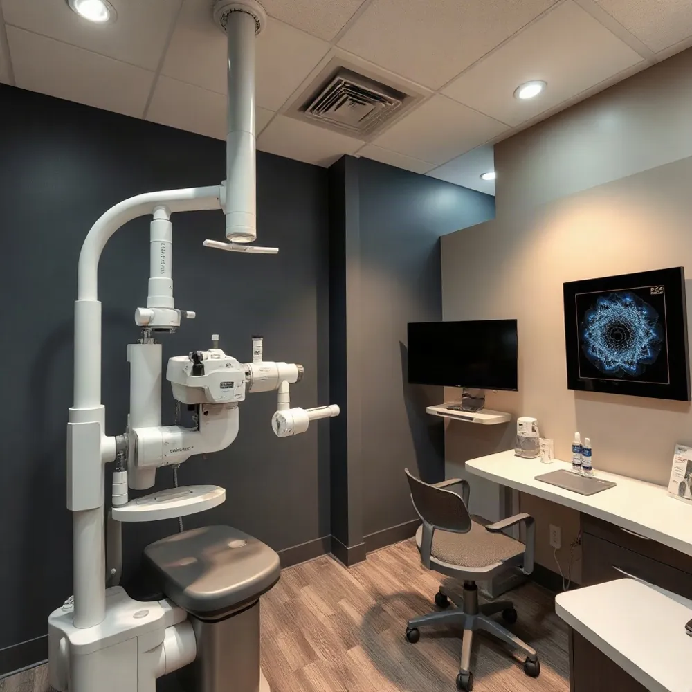
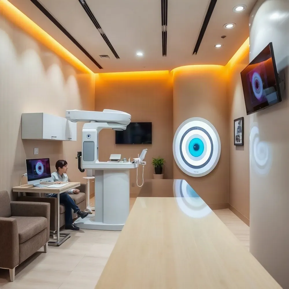
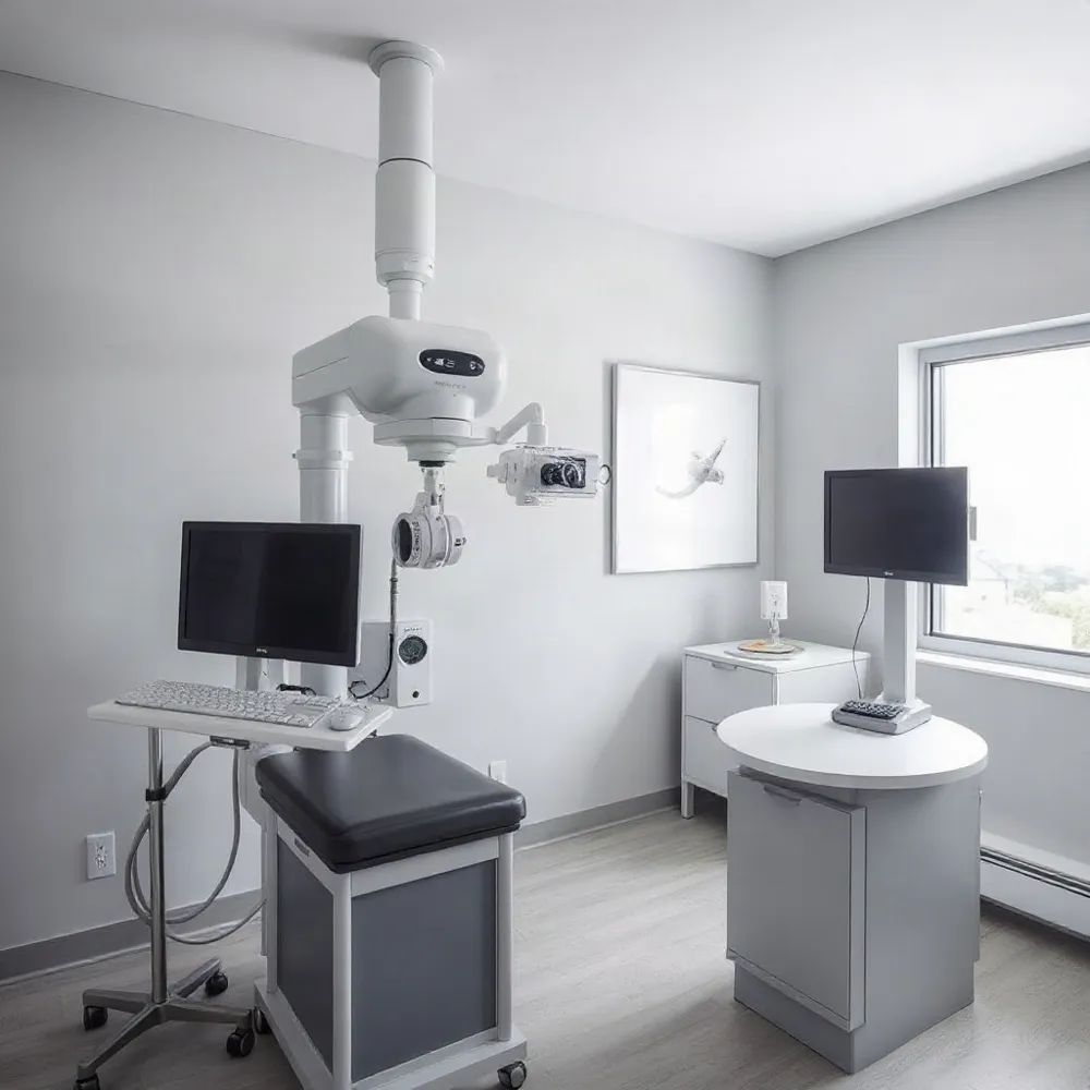
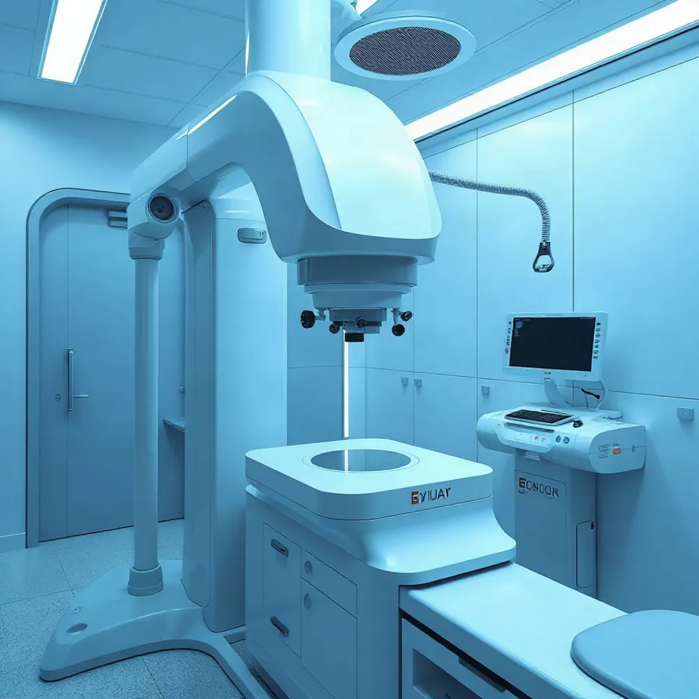
Advanced diagnostic technology is reshaping how eye care is delivered. Devices like the DRI OCT Triton provide detailed, cross-sectional images of the retina and deeper structures of the eye, allowing clinicians to detect conditions such as age-related macular degeneration and glaucoma earlier than ever before.
How DRI OCT Triton Works
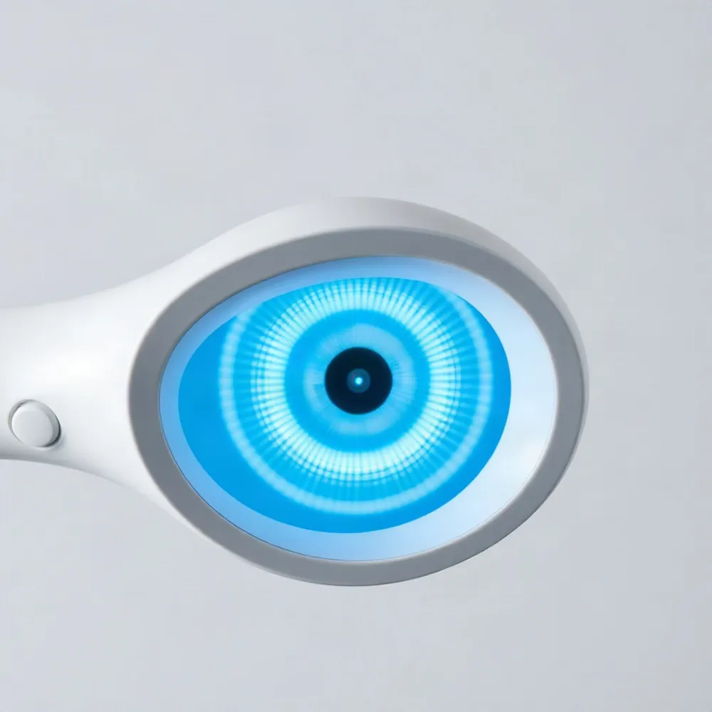 The Topcon DRI OCT Triton is an advanced diagnostic imaging platform that combines a deep-penetrating 1050nm wavelength Swept Source OCT (SS-OCT) with a scanning speed of 100,000 A-scans per second. This allows it to capture high-resolution, cross-sectional images from the vitreous down to the sclera, even through media opacities like cataracts or hemorrhages. The DRI OCT Triton was cleared by the FDA in 2018.
The device uses an invisible scan beam to reduce motion artifacts and help patients focus on the fixation target. This minimizes the need for rescans, making exams quicker and more comfortable. The built-in retinal camera and SMARTTrack eye tracking system work together to capture a dense data set of the retinal microvascular flow network.
Multimodal Imaging in One Scan
The DRI OCT Triton combines SS-OCT, OCT angiography (OCT-A), true color fundus photography, and fundus autofluorescence into a single device. Its wide-field scan (12mm x 9mm) covers both the optic nerve and macula in under two seconds — one reason retina specialist Daniel Lavinsky notes it provides almost all information needed for diagnosis and treatment decisions.
Proprietary algorithms like OCTARA enhance angiographic detection, while Smart Denoise reduces artifacts on OCT images. At Apple Eye Care in El Paso, TX, this technology is integrated into every comprehensive exam so patients can receive same-day follow-up or specialist referral without needing a hospital visit.
AI’s Growing Role in Diagnosis
 A 2025 survey found that 78% of ophthalmologists identified AI as the most transformative trend in their field, far ahead of other technologies. This enthusiasm is driven by real results: deep learning models, specifically convolutional neural networks, can now recognize patterns in imaging data for diabetic retinopathy, age-related macular degeneration (AMD), and glaucoma with accuracy equal to or exceeding human experts.
A study from University College London and Moorfields Eye Hospital compared an AI algorithm to human graders using 6,304 fundus images. The AI correctly identified glaucoma patients in 88-90% of cases, compared with 79-81% for humans. For AMD, AI-driven models go further by analyzing OCT images and clinical data to predict how patients will respond to treatments like anti-VEGF injections, helping clinicians decide who will benefit most and who may need alternative strategies.
AI models can also identify patients at high risk for rapid disease progression. This allows care teams to start aggressive treatment early, allocate resources more efficiently, and potentially reduce overall costs while improving patient outcomes.
Real-World AI Success Stories
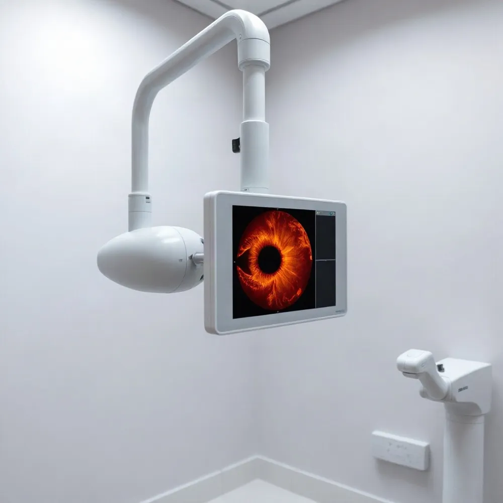 The first FDA-authorized autonomous AI system for diabetic retinopathy screening, IDx-DR (now LumineticsCore), received clearance in 2018. It analyzes retinal images without needing a specialist to interpret the results on-site, a milestone that opened the door for AI-powered screening in primary care settings.
A study at Johns Hopkins Children's Center demonstrated how such technology can close care gaps. Among youth with diabetes, 100% of participants completed an AI-driven camera exam versus only 22% who completed a traditional referral to an eye doctor within six months. The autonomous system takes four images of the eye without dilation and runs them through an algorithm to detect diabetic retinopathy on the spot.
Sutter Health expanded this model into primary care. Over a three-month pilot using AI-enhanced cameras, its six sites closed 235 diabetic eye exam gaps — with 92% of completed exams done via the AI system in primary care rather than by referral. By January 2025, Sutter scaled the technology to 28 practices, raising diabetic eye exam compliance from 62% to 68%, with a goal of reaching 72% by year-end.
For anterior segment diseases, the deep learning model CorneAI shows similar impact. In a study published in Scientific Reports, ophthalmologists using CorneAI support improved their diagnostic accuracy from 79.2% to 88.8%, with resident accuracy jumping from 75.6% to 86.2%. The model itself had an accuracy of just 86%, meaning it acted as a collaborative tool that raised clinicians' performance above its own baseline.
| Application | Technology | Impact |
|---|---|---|
| Diabetic retinopathy screening | IDx-DR (FDA cleared) | First autonomous AI system; used in >15,000 Medicare claims |
| Diabetic retinopathy (youth) | Autonomous camera + algorithm | 100% completion vs. 22% for traditional referrals (Johns Hopkins) |
| Diabetic eye exam compliance | AI-enhanced cameras in primary care | +6 percentage point compliance lift (Sutter Health) |
| Cataract & corneal diagnosis | CorneAI deep learning model | +9.6 percentage point accuracy improvement for ophthalmologists |
Expanding Access with AI-Enhanced Diagnostics
Traditional eye exams create significant barriers. Patients often need to schedule a separate visit with a specialist, undergo uncomfortable dilation drops that blur vision for hours, and manage transportation and time away from work. These hurdles lead to low screening rates.
A study by Johns Hopkins Children's Center highlights the stark difference: only 22% of youth with diabetes completed a traditional referral to an eye doctor within six months, while 100% completed an AI-driven exam that uses a retinal camera without dilation and delivers results in under 60 seconds.
Sutter Health's pilot program demonstrated how this approach scales. During a three-month trial at six primary care sites, the health system used AI-enhanced cameras to close 235 diabetic eye exam gaps. By January 2025, Sutter scaled the program to 28 primary care practices, raising overall diabetic eye exam compliance from 62% to 68%.
AI-powered screening tools are especially valuable for closing care gaps among racial and ethnic minorities and rural populations who face higher rates of diabetic eye disease and lower access to regular screenings. The Johns Hopkins study found no statistical differences in follow-through based on race, gender, or socioeconomic status when using autonomous AI exams.
Apple Eye Care integrates these advanced diagnostics into every comprehensive exam at its El Paso practice. The clinic's DRI OCT Triton provides high-density OCT scans alongside wide-field retinal imaging, offering patients dilation-free screenings that can be reviewed immediately by Dr. Stephen Applebaum — without requiring a separate hospital referral.
Treating What’s Detected: AMD and Glaucoma Care
Early detection of age-related macular degeneration (AMD) and glaucoma is critical because these diseases often progress without noticeable symptoms. Glaucoma can cause up to 40% of optic nerve fiber loss before visual symptoms appear, making routine imaging with tools like the DRI OCT Triton essential. By using optical coherence tomography to capture cross-sectional images of the retina and optic nerve, eye care providers can identify subtle structural changes years before a patient notices vision problems.
For dry AMD, the standard treatment for over a decade has been antioxidant vitamins, but recent research is yielding promising new options. A major area of advancement involves therapies that target geographic atrophy, with some new drugs in late-stage clinical trials aiming to slow or halt the growth of these lesions. While not yet widely available, these emerging treatments offer hope for more effective management beyond vitamin therapy alone.
AI Guides Treatment Decisions
Artificial intelligence is increasingly used to predict how individual patients will respond to treatments. For AMD, AI models analyze OCT images alongside clinical data to determine who will benefit most from anti-VEGF injections or other therapies, allowing doctors to tailor care plans more effectively. This data-driven approach helps avoid trial-and-error prescribing and ensures resources are directed toward patients who need them most.
AI Cannot Cure Glaucoma
While AI cannot cure glaucoma, it transforms how the disease is diagnosed and managed. Algorithms can detect subtle changes in the optic nerve head and retinal nerve fiber layer on fundus photographs or OCT scans with consistency that may exceed manual human review. Researchers are also developing smartphone-based AI tools to make screening accessible in underserved areas, but experts emphasize that AI assists specialists by improving diagnostic accuracy rather than replacing comprehensive medical care.
AI as a Collaborative Partner, Not a Replacement
The concern that AI will replace eye care professionals is widespread, but the evidence points in a different direction. A 2025 study on CorneAI, a deep learning model for diagnosing cataracts and corneal diseases, showed that when 40 ophthalmologists used AI support, their diagnostic accuracy rose from 79.2% to 88.8%. The AI likely values each of nine categories per image, helping clinicians narrow down possibilities—but the final diagnosis still rests with the doctor.
At Apple Eye Care in El Paso, TX, AI plays a similar supporting role. The practice uses AI algorithms to automatically compare scans against large datasets of normal and diseased eyes and flag suspicious regions. But Dr. Stephen Applebaum and his team review each Triton’s high-density scan to confirm findings. The goal is to use AI as a tool that enhances clinical judgment, not overrides it.
Where AI Falls Short
AI systems can fail when applied outside the specific conditions they were trained on—such as a different camera model, patient ethnicity, or disease severity. Their accuracy depends heavily on the quality and diversity of the training data. Many ophthalmic AI tools remain assistive rather than autonomous and should always be interpreted alongside the patient’s history and the doctor’s exam.
By handling time-consuming tasks like image analysis and preliminary screening, this technology frees clinicians to focus on patient care, education, and treatment planning. The doctor still reviews all results, considers individual health factors, and determines next steps.
Transforming Drug Development and Research
AI's impact on ophthalmology extends beyond the clinic into drug development and clinical research. In early-stage drug discovery, AI can cut development time by over 60% and improve predictions of a protein-based drug's efficacy, safety, and manufacturability. Large language models can also generate novel molecular structures and predict drug-target interactions, accelerating the identification of promising compounds.
Clinical trial recruitment is another area where AI provides value. Approximately 80% of trials fail to enroll on time, but AI algorithms can rapidly identify eligible patients from large datasets, reducing timelines and the number of participants needed. While shorter development cycles and reduced costs are clear benefits for new ophthalmic drugs, standardized regulations will be necessary to ensure safe and equitable implementation.
A Brighter Future for Eye Health
Advanced diagnostics and artificial intelligence are already reshaping how eye disease is detected, treated, and managed. The combination of swept-source OCT platforms like the DRI OCT Triton with AI-powered analysis allows clinicians to identify conditions such as glaucoma, diabetic retinopathy, and age-related macular degeneration earlier than ever before.
Apple Eye Care in El Paso integrates these technologies into every comprehensive exam, making advanced screening accessible without requiring a separate specialist visit. The practice uses the DRI OCT Triton to capture high-resolution cross-sectional images of the retina, optic nerve, and choroid in a single scan. AI algorithms then compare those scans against large datasets to flag suspicious regions for the doctor's review. This approach combines the speed of automated analysis with the clinical judgment of Dr. Stephen Applebaum and his team.
Innovation continues on the horizon. Wearable devices and IoT sensors could soon enable continuous eye health monitoring, alerting patients and providers to changes that require intervention. Predictive AI models are being developed to forecast disease progression, allowing earlier treatment adjustments. Teleophthalmology platforms already bring screenings to rural and underserved communities increasing access.
The future of eye care is proactive, personalized, and powered by technology – but guided by compassionate professionals who interpret results within each patient's full health picture.