The Evolution of Custom Contact Lens Fitting
Traditional assessment methods, such as manual keratometry, are limited by their ability to measure only the central 3mm of the cornea, according to the American Academy of Ophthalmology. This approach often fails to capture the full surface profile required for specialized contact lenses.
Modern corneal topography uses advanced light-based scanning to map thousands of points, providing a detailed surface blueprint. At Apple Eye Care, this shift toward high-resolution diagnostic data allows for personalized plans that improve patient outcomes. By integrating tools like the DRI OCT Triton, the practice achieves a level of precision that older methods simply cannot match.
Individual topographical mapping is essential for patients with unique corneal shapes or conditions like keratoconus. Per the Review of Contact Lenses, these detailed maps are critical for selecting the correct lens diameter and base curve, ensuring the stability and comfort that one-size-fits-all options lack. Reliable data remains the foundation for long-term vision health and successful lens performance.
Understanding Corneal Topography and Its Clinical Importance
Corneal topography is a quick, non-invasive diagnostic test that creates detailed, color-coded maps of the cornea’s surface curvature and elevation. By acting as a topographic map for the eye, it allows our team at Apple Eye Care to identify subtle surface irregularities that are invisible during a standard examination. This test is essential for diagnosing and monitoring conditions like keratoconus, astigmatism, and corneal scarring. Furthermore, it serves as a critical tool for surgical planning, including LASIK, cataract, and transplant procedures, to ensure optimal outcomes per American Academy of Ophthalmology standards. We also use these precise measurements to customize specialty contact lenses, ensuring a comfortable and accurate fit tailored specifically to your eye's unique shape.
Visualizing the Cornea
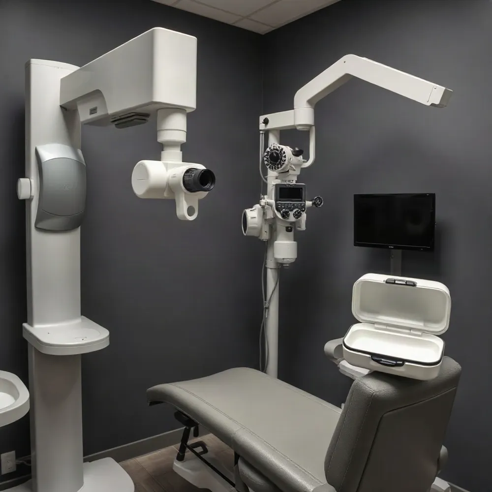
The diagnostic process involves a patient resting their head in a stable position while looking into a bowl-shaped device. This instrument projects light patterns onto the eye, capturing thousands of data points to generate a map of the surface terrain. Unlike traditional manual keratometry, which only measures the central 3mm of the cornea, topography provides a comprehensive view of the entire anterior surface. At Apple Eye Care, we integrate this data to guide the selection of gas-permeable or scleral lenses, ensuring that the final fit addresses individual variations in corneal height and curvature.
| Map Type | Clinical Purpose | Key Advantage |
|---|---|---|
| Axial | Curvature | Determines base curves |
| Elevation | Height | Visualizes lens fit |
| Tangential | Peripheral | Detects irregularities |
The Patient Experience During Corneal Mapping
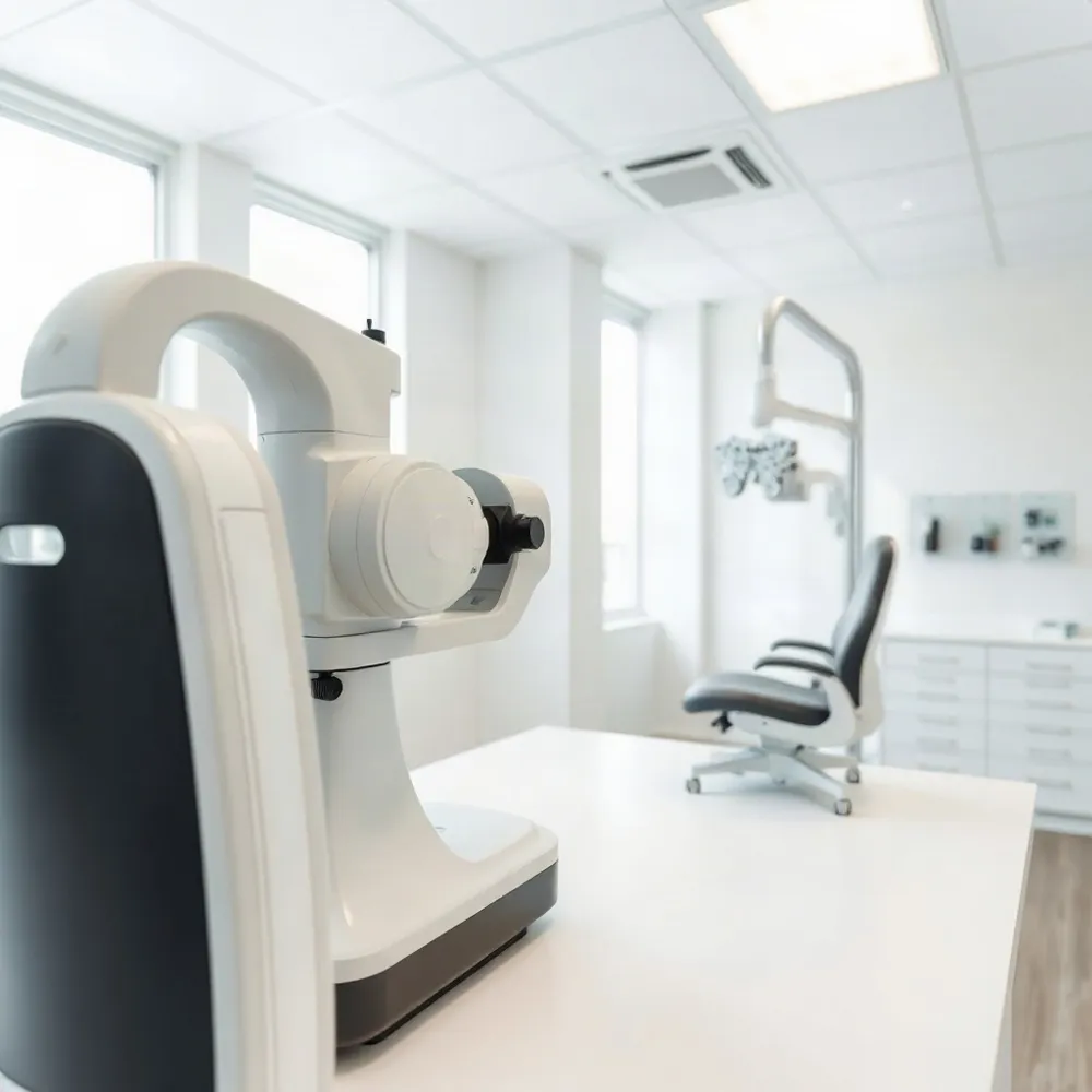 Understanding what to expect during a diagnostic evaluation helps alleviate common concerns about eye health assessments. Corneal topography is a quick, non-invasive, and completely painless diagnostic test used to create a detailed map of your cornea’s surface. During the procedure, you simply rest your chin and forehead against the device while looking toward a target light inside the machine. As you hold steady, a camera captures images of your eye in a matter of seconds without any part of the device ever touching your eye according to the American Academy of Ophthalmology.
Preparation is a vital part of obtaining an accurate corneal topography result. Because contact lenses can cause temporary shifts in corneal shape known as corneal warpage, patients are often required to discontinue lens wear for a specific period before their appointment as noted by the NCBI. This pause ensures the cornea returns to its natural resting state, allowing for measurements that reflect your true eye structure rather than temporary distortion.
At Apple Eye Care, we integrate these high-resolution images with our clinical expertise to provide personalized care tailored to the unique shape of your eye. This process allows our team to identify subtle elevation changes, diagnose conditions like astigmatism or keratoconus, and assist in planning refractive or cataract surgeries. By providing these precise, color-coded maps, we ensure that every treatment plan is grounded in exact diagnostic data, moving away from generic models to focus on individual visual demands.
Distinguishing Between Topography and Advanced Tomography
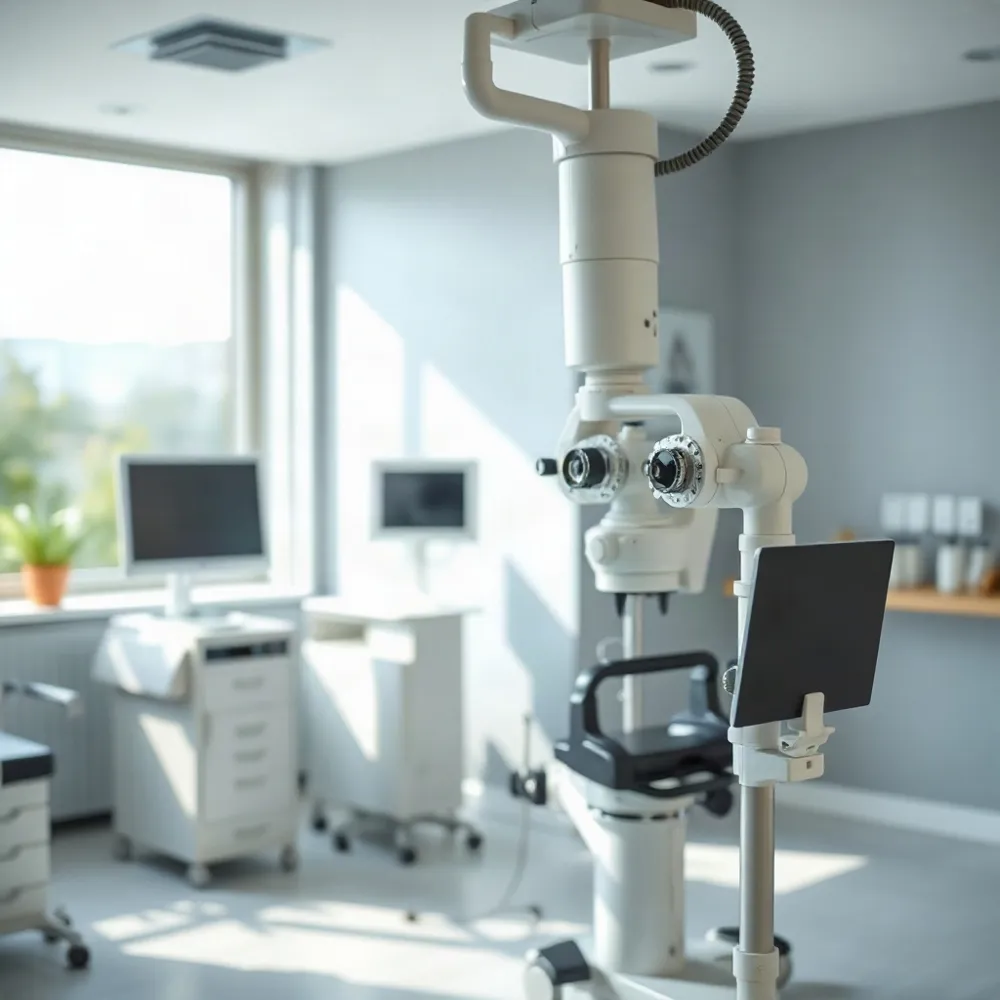 While often conflated, the terms topography and tomography refer to distinct diagnostic technologies. Standard corneal topography utilizes a Placido-based reflection to create a two-dimensional map of the anterior corneal surface. This process is effective for general contact lens fitting, but because it relies on the reflection of light off the tear film, it cannot measure the posterior surface or the overall thickness of the tissue, per NECO.
Advanced systems like the Pentacam perform corneal tomography, a method that captures a comprehensive three-dimensional structure of the eye. By using Scheimpflug imaging—a technique involving a rotating camera that generates cross-sectional images—these systems map both the anterior and posterior surfaces. This allows for precise measurement of corneal thickness and elevation, which surface-level mapping might overlook.
At Apple Eye Care, we recognize that these detailed structural maps are essential for identifying early indicators of conditions like keratoconus or ectasia. Unlike the traditional curvature analysis used by many competitors, our approach integrates advanced diagnostic data to inform custom lens design. By mapping the full 3D structure of the eye, we ensure a higher degree of precision for patients with complex corneal shapes, moving beyond the limitations of surface-only assessments.
Interpreting Results and Understanding Normal Corneal Anatomy
Corneal topography maps utilize a color-coded system to visualize the surface shape and elevation of your eye, where warm colors such as red and orange typically indicate steeper or thinner areas, while cool colors like blue and green represent flatter or thicker regions my.clevelandclinic.org/health/diagnostics/24819-corneal-topography. Clinicians review several maps to build a complete profile, including the axial map for curvature and the pachymetry map for corneal thickness my.clevelandclinic.org/health/diagnostics/24819-corneal-topography. A normal result generally displays a symmetric, bowtie-shaped pattern on the axial map, which signifies regular astigmatism markennovy.com/blog/how-to-read-corneal-topography-for-contact-lens-fitting/.
How should patients interpret corneal topography maps and what constitutes a normal result?
A healthy cornea typically shows gradual, progressive flattening from the center toward the periphery, with specific elevation and thickness values that fall within expected clinical ranges www.reviewofcontactlenses.com/article/putting-it-on-the-map-fitting-rigid-lenses-using-corneal-topography. While these maps are highly detailed, they are just one piece of the diagnostic puzzle. At Apple Eye Care, Dr. Stephen Applebaum integrates these topographical findings with clinical expertise to monitor eye health and determine candidacy for specialized lens designs.
Because raw data can be complex, our team at Apple Eye Care conducts a thorough review of your specific measurements during your follow-up visit. Unlike generic screening tools used at other practices, our personalized approach involves explaining these visual maps to ensure you feel confident and informed about your eye care plan. If your results show deviations from normal ranges, we use this precise data to customize contact lenses that account for your unique corneal structure, providing sharper vision compared to standard, one-size-fits-all alternatives.
Leveraging Topography for Optimized Contact Lens Fitting
 Eye care providers utilize corneal topography to transcend the limitations of traditional keratometry, which only measures the central few millimeters of the eye. By projecting light patterns and measuring their reflection, this technology generates comprehensive, color-coded maps of the cornea's curvature, asphericity, and symmetry. At Apple Eye Care, this data serves as a blueprint for custom contact lens selection, ensuring the final lens profile accounts for peripheral irregularities that standard tools often miss.
How do eye care providers utilize corneal topography for contact lens fitting?
Corneal topography utilizes advanced light-based scanning to generate detailed, color-coded maps of the eye’s surface, allowing eye care providers to precisely measure central curvature, symmetry, and asphericity. By analyzing these maps, practitioners can determine the ideal lens design, diameter, and base curve, ensuring the contact lens aligns correctly with the patient's unique corneal profile. This technology is especially vital for identifying irregularities such as astigmatism or keratoconus, which often require specialized lenses for optimal comfort and visual performance. Additionally, providers use these maps to assess how the cornea deviates from a perfect sphere, helping them select the most stable and centered fit for each individual. By integrating these measurements, doctors can proactively troubleshoot potential fit issues and provide personalized, accurate vision correction.
Beyond selecting the initial lens, these maps are essential for troubleshooting stability. If a patient experiences fluctuating vision, topographical data helps identify if the lens is decentered or if the corneal surface has developed warpage due to improper fit. While some practices rely on generic fitting models, personalized care relies on comparing baseline maps to current ocular health data. This proactive approach ensures that patients maintain consistent vision quality while minimizing the risk of complications from long-term lens wear.
Essential Standards and Myths in Vision Maintenance
What are the recommended guidelines for digital eye strain prevention, such as the 20-20-20 rule?
To help manage digital eye strain, many eye care professionals recommend the 20-20-20 rule: every 20 minutes, take a 20-second break to look at something at least 20 feet away. While this practice is widely promoted as a helpful way to reset your focus and reduce fatigue, clinical research on its effectiveness remains limited. Beyond this technique, digital eye strain can cause symptoms such as dryness, blurred vision, headaches, and light sensitivity, often stemming from prolonged screen use. If these symptoms persist despite taking regular breaks, a professional evaluation is necessary. Apple Eye Care helps determine if your discomfort is related to digital strain or other underlying issues, ensuring you receive personalized care for your long-term eye health.
Can comprehensive eye exams be effectively performed via telehealth or remote platforms?
While telemedicine and teleophthalmology offer valuable tools for remote consultations, they cannot replace the necessity of a comprehensive in-person eye exam. A truly comprehensive exam requires physical contact and specialized diagnostic instrumentation, such as the DRI OCT Triton used at Apple Eye Care, to perform a thorough assessment of internal ocular health and structural integrity. Remote platforms are effective for managing follow-up care, discussing symptoms, or assessing external issues, but they lack the ability to perform high-resolution, hands-on diagnostics like intraocular pressure checks or slit-lamp biomicroscopy. Consequently, telehealth should be viewed as a vital supplement to, rather than a substitute for, the clinical accuracy provided during an in-office visit. Regular, face-to-face examinations remain the gold standard for early detection and long-term management of conditions like glaucoma, cataracts, and other sight-threatening diseases.
There is conflicting information regarding the '3:1:1' and '4:1' rules for contact lens wear; what are the established safety guidelines?
There are no medically recognized standards known as the 3:1:1 or 4:1 rules for contact lens wear. Contact lens safety is governed by strict hygiene protocols established by the FDA and professional eye care associations, which emphasize that all lenses must be prescribed and fitted by a doctor of optometry. Key safety guidelines include washing your hands before touching your eyes, cleaning and disinfecting lenses daily as directed, and never using tap water or saliva for cleaning. It is essential to strictly adhere to your specific wear schedule, dispose of lenses as instructed, and never sleep in them unless specifically cleared by your eye doctor. If you experience any pain, redness, or blurred vision, you should remove your lenses immediately and contact your optometrist for professional guidance.
Prioritizing Your Long-Term Vision Health
Achieving clear, comfortable vision depends on more than a single prescription. At Apple Eye Care, patients receive personalized care plans that integrate high-resolution data from tools like the DRI OCT Triton to monitor subtle changes over time. By shifting away from one-size-fits-all models, this tailored approach reduces emergency eye-care visits by 15% to 20% while significantly boosting patient satisfaction.
Ongoing monitoring of your corneal shape is essential, especially if you wear contact lenses. Routine scans help detect early signs of corneal warpage or compression, allowing for timely adjustments that preserve long-term ocular health. If you are interested in a professional consultation to discuss your specific needs, visit www.appleeyecare.com to schedule an appointment with Dr. Stephen Applebaum and his dedicated team.