The Evolution of Modern Vision Care
Modern optometry is defined by the seamless marriage of high-resolution diagnostic precision and patient-centered clinical expertise. At Apple Eye Care, the commitment to incorporating advanced technology ensures that every screening moves beyond surface-level evaluation into a detailed, microscopic analysis of ocular health. By utilizing tools like the DRI OCT Triton, the practice achieves a level of diagnostic clarity that standard systems often overlook.
While many providers rely on traditional imaging, the DRI OCT Triton employs swept-source technology to visualize deeper eye structures, such as the choroid and sclera, even through media opacities like cataracts. This technical capability permits earlier detection of conditions that threaten long-term vision, as noted by studies highlighting how optical coherence tomography provides objective quantification of disease damage far sooner than functional testing alone.
Technological adoption remains an extension of, rather than a replacement for, clinical judgment. In an era where artificial intelligence continuously improves triage and diagnostic consistency, the compassionate care provided at Apple Eye Care ensures that every data point translates into a personalized management plan. This balanced approach protects patient autonomy while delivering the benefits of modern innovation.
Understanding Optical Coherence Tomography Technology
![]() An Optical Coherence Tomography (OCT) eye test is a non-invasive, advanced imaging technology that uses light waves to create detailed, cross-section and 3D images of your retina and optic nerve. Unlike standard eye exams, OCT reveals each distinct layer of the retina in microscopic detail, allowing for precise measurement of thickness and changes over time. It is performed to help diagnose, treat, and manage leading causes of vision loss, including glaucoma, age-related macular degeneration, diabetic retinopathy, and macular holes. The test is quick and painless, typically taking less than 10 minutes, and can detect signs of disease often years before symptoms appear. At Apple Eye Care, this advanced imaging allows our team to provide highly personalized care for conditions like glaucoma and retinal disease.
An Optical Coherence Tomography (OCT) eye test is a non-invasive, advanced imaging technology that uses light waves to create detailed, cross-section and 3D images of your retina and optic nerve. Unlike standard eye exams, OCT reveals each distinct layer of the retina in microscopic detail, allowing for precise measurement of thickness and changes over time. It is performed to help diagnose, treat, and manage leading causes of vision loss, including glaucoma, age-related macular degeneration, diabetic retinopathy, and macular holes. The test is quick and painless, typically taking less than 10 minutes, and can detect signs of disease often years before symptoms appear. At Apple Eye Care, this advanced imaging allows our team to provide highly personalized care for conditions like glaucoma and retinal disease.
The procedure functions by directing infrared light into the eye. These waves reflect off the retinal structures, and the device captures the data to construct a map of the internal anatomy. This process mirrors the mechanics of ultrasound imaging, yet it provides superior resolution by utilizing light instead of sound to visualize structures often measured in microns. Because the scan is non-contact, it ensures maximum comfort while gathering the precise data needed for clinical decision-making.
Clinical Applications for Early Detection
- Detection of microscopic retinal thinning linked to glaucoma progression.
- Precision monitoring of macular edema in diabetic patients.
- Longitudinal comparison of structural anatomy to halt preventable sight loss.
- Visualization of vitreoretinal relationships for advanced surgical planning.
Beyond routine checks, this technology is a cornerstone of proactive eye health. By capturing high-definition cross-sections, clinicians can assess structural changes long before a patient experiences noticeable vision loss. At Apple Eye Care, we integrate these findings to monitor stable patients and quickly triage those showing early signs of pathology. This objective data empowers our team to deliver consistent care that prioritizes your visual longevity.
Patient Experience During Advanced OCT Screenings
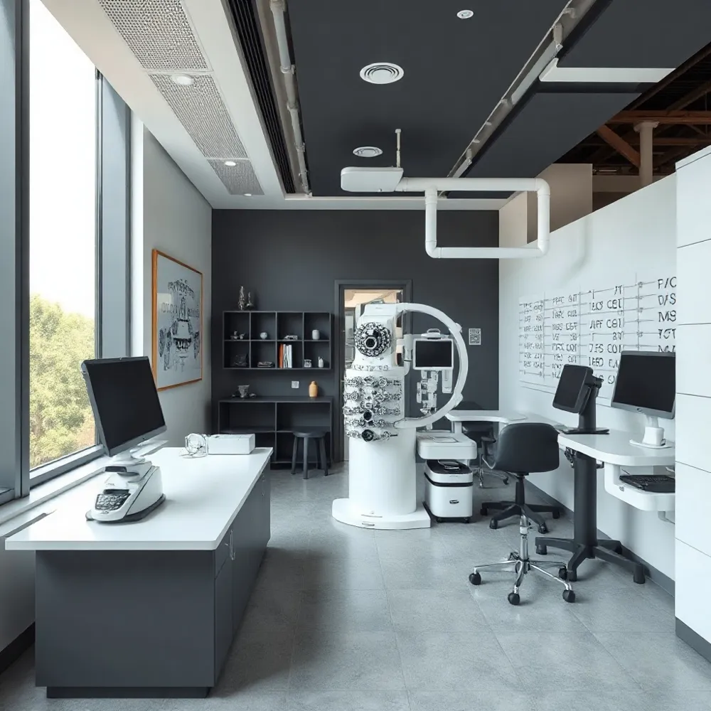
![]() Achieving diagnostic precision begins with patient comfort. When you visit Apple Eye Care for advanced diagnostics, our team utilizes the DRI OCT Triton to streamline your screening. Unlike traditional testing methods that may involve invasive techniques or prolonged discomfort, this technology is entirely non-contact. You simply rest your chin on a supportive cradle and focus on a target inside the device for a few seconds.
Achieving diagnostic precision begins with patient comfort. When you visit Apple Eye Care for advanced diagnostics, our team utilizes the DRI OCT Triton to streamline your screening. Unlike traditional testing methods that may involve invasive techniques or prolonged discomfort, this technology is entirely non-contact. You simply rest your chin on a supportive cradle and focus on a target inside the device for a few seconds.
What can patients expect during an OCT eye test procedure? Most screenings take fewer than 10 minutes to complete, per OHSU Casey Eye Institute research. Because the system captures images using light waves at a high speed of 100,000 A-scans per second, as noted in Retina Specialist, the risk of motion artifacts from blinks or involuntary eye movement is significantly reduced. This efficiency allows our specialists to obtain high-resolution data quickly and comfortably.
By integrating such sophisticated technology into our routine practice, we ensure that every patient receives a comprehensive analysis without the stress often associated with complex screenings. The non-invasive nature of the Optical Coherence Tomography process ensures you remain relaxed while we map the delicate structures of your retina and optic nerve. This focus on a seamless, professional experience remains a standard hallmark of the care you will find at our clinic.
Precision Management of Glaucoma with DRI OCT Triton
![]() Effective glaucoma management demands a shift from reactive care to proactive, data-driven monitoring. At Apple Eye Care, we rely on the DRI OCT Triton to elevate this process. Unlike standard systems, this instrument utilizes swept-source technology to achieve a 1,050-nm light source, allowing deeper penetration through internal ocular tissues. This capability is essential for visualizing the retina, choroid, and sclera with high clarity, even in patients who have media opacities like cataracts that often obscure diagnostic views in lesser devices.
Effective glaucoma management demands a shift from reactive care to proactive, data-driven monitoring. At Apple Eye Care, we rely on the DRI OCT Triton to elevate this process. Unlike standard systems, this instrument utilizes swept-source technology to achieve a 1,050-nm light source, allowing deeper penetration through internal ocular tissues. This capability is essential for visualizing the retina, choroid, and sclera with high clarity, even in patients who have media opacities like cataracts that often obscure diagnostic views in lesser devices.
How are modern diagnostic tools like the DRI OCT Triton utilized in glaucoma management? The DRI OCT Triton utilizes swept-source technology to provide deep, high-clarity visualization of ocular structures. This precision is vital for the early detection and ongoing management of glaucoma, allowing doctors at Apple Eye Care to track subtle changes in the optic nerve and retinal nerve fiber layer over time. By capturing 100,000 A-scans per second, the system minimizes artifacts caused by natural eye movement and creates an expansive, high-resolution dataset.
Consistent baseline monitoring is the cornerstone of preserving vision, as glaucoma often progresses without identifiable symptoms. The Triton allows for multimodal imaging, combining fundus photography and objective layer measurements into a single capture. This provides an integrated view of retinal nerve fiber layer health and optic disc integrity. By automatically mapping findings and comparing them against established clinical databases, practitioners can isolate even minor anatomical shifts that warrant intervention, ensuring that care plans are adjusted long before functional loss occurs.
The Transformative Influence of Artificial Intelligence in Optometry
Artificial intelligence is changing modern optometry by acting as a powerful clinical and operational tool that empowers providers. By integrating sophisticated data processing into routine eye exams, eye care teams can now bridge service gaps and enhance the accuracy of diagnostic interpretations. While some clinicians initially expressed caution, roughly 72% of optometrists now believe that these tools meaningfully improve their daily practice according to a 2022 Journal of Optometry study.
How is artificial intelligence (AI) being integrated into modern optometry and patient care?
Advanced deep learning models are currently able to analyze high-resolution imagery to detect subtle biomarkers for conditions like diabetic retinopathy, glaucoma, and age-related macular degeneration with accuracy that often rivals human experts per research published in the National Library of Medicine. By utilizing Convolutional Neural Networks, these systems can identify complex patterns in ocular scans, allowing for earlier intervention before visible symptoms manifest.
Beyond the diagnostic suite, practice management is undergoing a shift toward increased efficiency. AI-supported tools can alleviate administrative burdens, such as automated charting or scheduling, which are primary factors in reducing clinical fatigue as noted by the American Optometric Association.
At Apple Eye Care in El Paso, the priority remains a human-centric approach that blends precision technology with compassionate care. Unlike autonomous systems that risk isolating patients from their clinicians, the integration of intelligent diagnostics here is designed to augment the expertise of Dr. Stephen Applebaum. By automating data-heavy analytical tasks, our practice ensures that more time is directed toward meaningful patient interaction, managing chronic conditions, and fostering long-term wellness for every family we serve.
Advancing Outcomes in Modern Glaucoma Care
Effective glaucoma management is shifting from a reactive reliance on daily topical medications toward a proactive interventional model. Traditional approaches often face challenges with high patient nonadherence and intraocular pressure fluctuations that go undetected by periodic office visits, according to research on early diagnostics and interventional glaucoma. At Apple Eye Care, we enhance this clinical oversight by using the DRI OCT Triton to capture high-resolution, objective data that identifies progression far sooner than functional visual field tests alone.
What is the current state of glaucoma research and treatment outcomes for patients?
Glaucoma management has evolved significantly with intensive research into neuroprotection, innovative surgical techniques, and advanced monitoring devices. While the condition remains a challenge, modern diagnosis and personalized treatment plans offer patients a promising path to preserving vision. By integrating home-based rebound tonometers like the iCare HOME, clinicians can now track pressure patterns outside of clinical settings to bridge gaps in patient care. This comprehensive data allows for personalized interventions, such as minimally invasive glaucoma surgery, tailored to the individual patient profile.
Future research is moving toward identifying risk markers through genetic testing panels and mitochondrial monitoring. Apple Eye Care supports these advancements by ensuring that the objective metrics captured by our imaging technology directly inform patient treatment strategies, helping to mitigate the risks of sight-threatening vision loss. By focusing on predictive care rather than symptomatic response, providers can better manage ocular health for the long term.
A Future Defined by Advanced Clinical Care
Modern eye care is reaching a turning point where high-tech diagnostic capability meets the irreplaceable human touch. Technologies like the DRI OCT Triton allow providers to see deep into the eye with unprecedented clarity, catching microscopic changes long before they manifest as vision loss, according to OHSU Casey Eye Institute. While these tools offer deep precision and predictive modeling, they function best as an extension of the clinician rather than a substitute for professional judgment.
At Apple Eye Care, we integrate these advanced diagnostics to serve our El Paso community with a standard of precision rarely seen in private practice. We prioritize a care model that values your time and health, pairing sophisticated OCT imaging with Dr. Stephen Applebaum's expertise to ensure your specific needs remain the primary focus. By using these systems as a foundation, we move away from reactive treatments and toward consistent, long-term wellness planning.
We invite you to experience the future of eye health locally. Whether you are managing chronic conditions or simply maintaining your vision, our team provides the compassionate environment necessary to navigate your health confidently. Schedule your comprehensive exam at www.appleeyecare.com to see how state-of-the-art technology can protect your sight for years to come.