Seeing Beyond the Eye Chart
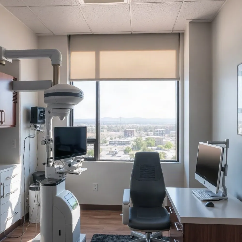
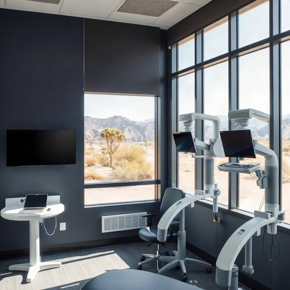
A comprehensive eye exam goes beyond reading letters on a chart. It can combine a conversation about vision and health, familiar vision tests, and an examination of the eyes. At Apple Eye Care in El Paso, advanced diagnostics, including DRI OCT Triton imaging, add another source of information for the clinician.
Different tools answer different questions. Wavefront testing assesses optical imperfections that may affect how light travels through the eye, while optical coherence tomography creates detailed images of eye structures. Artificial intelligence can assist with image analysis, but research describes it as support for clinical review, not a replacement for it.
No single test is right for everyone. The clinician considers a patient’s symptoms, history, and examination findings when deciding which tests may help. Results then contribute to a broader assessment and discussion of care.
The Foundation of a Comprehensive Eye Exam
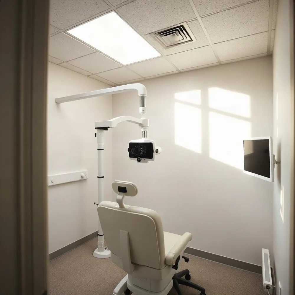 A comprehensive eye exam is a sequence of checks, not a single test. The optometrist chooses what to assess based on a patient’s age, symptoms, health history, and initial findings. At Apple Eye Care in El Paso, that evaluation can be considered alongside the practice’s advanced diagnostics, including DRI OCT Triton imaging when closer assessment is appropriate.
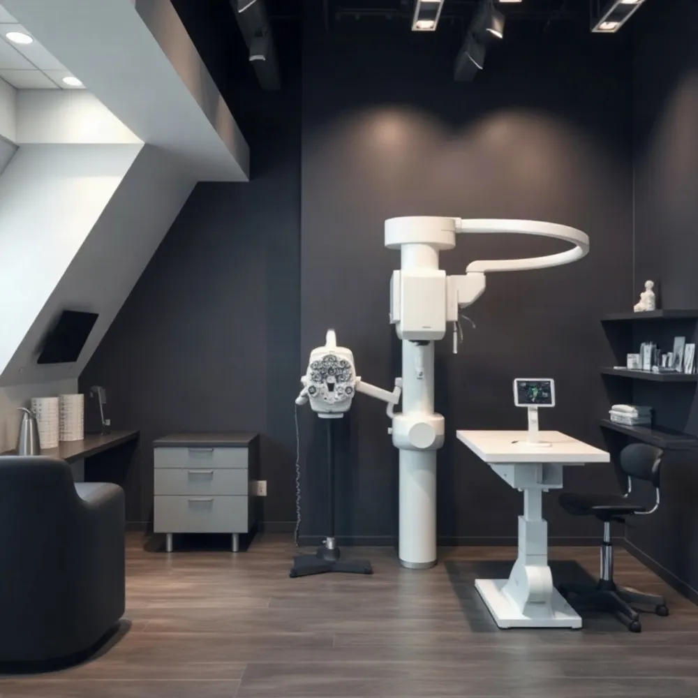
Visual acuity testing with a Snellen chart checks how clearly each eye sees at a distance. An autorefractor or retinoscope can estimate the prescription, then the optometrist refines it by comparing lens choices through a phoropter. The patient’s responses help determine which correction gives the clearest view.
A slit lamp provides a magnified view of structures at the front of the eye, while an ophthalmoscope helps the doctor examine the back. Dilating drops may widen the pupils to make more of the retina and other internal structures visible. The doctor may also measure eye pressure as one part of glaucoma screening, rather than treating that reading alone as a diagnosis.
Visual-field testing maps peripheral vision and can reveal areas a patient may not notice are missing. Depending on symptoms, risk factors, or findings, the optometrist may recommend imaging or other tests for a closer look. For example, optical coherence tomography is an established subject in ophthalmic diagnostic research, but imaging supports the clinical examination rather than replacing it (OCT research in ophthalmology).
Mapping the Eye’s Optical Imperfections
Wavefront analysis, also called aberrometry, measures how light travels through the eye’s focusing system. The test records distortions in the returning light and turns them into a map of optical imperfections, offering information beyond a standard prescription check. Wavefront testing is one way clinicians assess these optical differences.
The map can show lower-order errors, including nearsightedness, farsightedness, and astigmatism, as well as higher-order aberrations. These subtler irregularities may contribute to symptoms such as glare or halos, though a measured aberration does not necessarily cause a problem or require treatment.
Wavefront analysis and corneal topography describe different aspects of vision. Wavefront analysis assesses how light passes through the eye’s optical system, while topography maps the shape and curvature of the cornea’s front surface. An eye-care professional can decide whether either measurement, or both, is useful for a particular concern.
Results may help inform individualized vision-correction planning, but they do not guarantee a specific result or determine on their own whether someone is suitable for surgery. Tear-film changes, including those associated with dry eye, can also affect measurements. At Apple Eye Care, advanced diagnostics such as the DRI OCT Triton support eye-health assessment; any test findings still need to be interpreted alongside symptoms and a clinical examination.
What Retinal Imaging Can Reveal
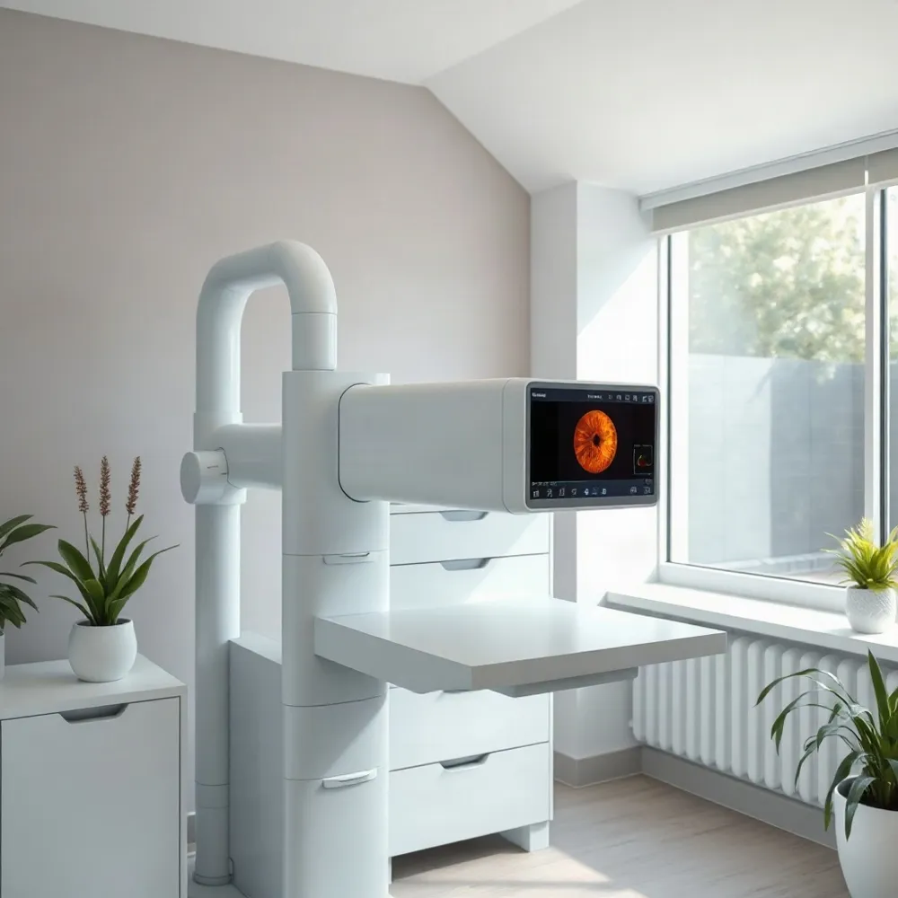
What is an OCT scan, and how does it differ from a retinal photograph?
Optical coherence tomography (OCT) is a noninvasive scan that uses reflected light to create detailed cross-sectional images of retinal and optic nerve layers. It lets an eye doctor examine structures beneath the retinal surface. A retinal photograph, by contrast, records a two-dimensional view of the retina’s appearance. The tests provide different information and may be used separately or together, depending on a patient’s eye health and risk factors. OCT is also an established subject of ophthalmic diagnostic research.
Why might an optometrist recommend an OCT scan?
An optometrist may recommend OCT to investigate unexplained vision changes, examine retinal or optic nerve structures more closely, or monitor changes associated with glaucoma, macular degeneration, or diabetic retinal disease. It can support early assessment and follow-up, but it is not necessary for every patient. The recommendation depends on symptoms, examination findings, and risk factors, and the images must be interpreted in context.
At Apple Eye Care in El Paso, the DRI OCT Triton is part of the practice’s advanced diagnostic services. The system combines swept-source OCT with fundus imaging. A 2018 report on the DRI OCT Triton describes its multimodal imaging design; available functions depend on the device configuration.
What can the DRI OCT Triton help an eye doctor examine?
Depending on its configuration, the system can help clinicians view structures such as the retina, vitreous, and choroid, with options that may include OCT angiography or color fundus imaging. Apple Eye Care’s imaging supports a broader eye-health assessment, rather than serving as a diagnosis on its own. An eye examination and other relevant tests help the clinician decide what the images mean and whether follow-up or care is needed.
Artificial Intelligence as Clinical Support
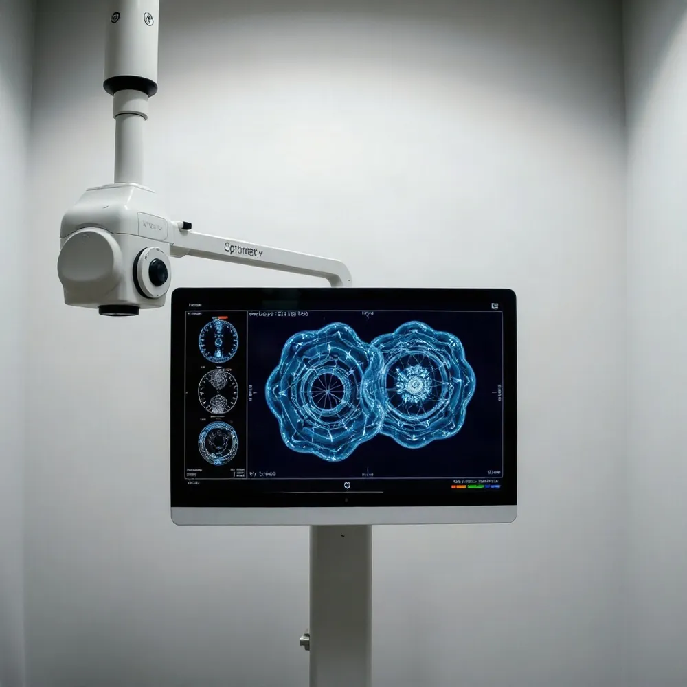 Artificial intelligence can help clinicians review eye images by identifying patterns and flagging areas that may need closer attention. In some health settings, AI systems also combine images with other patient information to help prioritize cases. These tools support review rather than establishing a diagnosis on their own.
A specific example is IDx-DR, which the FDA approved in 2018 as an autonomous tool to detect diabetic retinopathy without clinician input. That approval applies to this particular system and use; it does not mean that every AI tool for eye care is autonomous, approved, or used at Apple Eye Care.
AI-assisted analysis may help make image review faster and more consistent, and may draw attention to possible findings earlier. These are potential benefits, not guaranteed results for every system or patient. A flag from an algorithm is not the same as a confirmed condition, and the findings need to be considered alongside the patient’s history and examination.
At Apple Eye Care in El Paso, advanced diagnostics such as the DRI OCT Triton provide imaging that an eye-care professional can interpret in context. AI can inform clinical decisions, but it does not broadly replace the clinician’s judgment or the need for appropriate follow-up.
Technology Needs Context and Care
Different diagnostic tools answer different questions. Wavefront analysis maps optical imperfections that can affect how light is focused, while optical coherence tomography (OCT) creates cross-sectional images of eye structures. Artificial intelligence (AI), by contrast, analyzes data for patterns that may warrant closer review. At Apple Eye Care, advanced diagnostics such as the DRI OCT Triton contribute imaging information that an eye-care professional interprets alongside the broader examination.
AI adoption also raises practical concerns. Patient privacy, integration with older electronic health record systems, inconsistent development standards, and questions about accountability can affect how safely these tools are used. Responsible use depends on transparency about a system’s role and limits, careful attention to safety, and collaboration among clinicians, developers, and regulators. AI can support clinical review, but its output should not be treated as a diagnosis on its own.
When an eye doctor recommends a test, ask what it measures, why it may be useful in your situation, and how its findings fit with the rest of your evaluation. Understanding the purpose of a scan or analysis can help make the results more meaningful and keep decisions grounded in your individual needs.
A More Informed View of Eye Health
Modern tools can add useful detail to an eye-care evaluation, but their findings matter most when a clinician interprets them alongside a patient’s symptoms, history, and needs. At Apple Eye Care in El Paso, advanced diagnostics such as the DRI OCT Triton support that broader clinical assessment.
No single scan, optical map, or algorithm answers every question. Ask your eye-care professional which tests may help clarify your concerns and how the results fit into your care plan.