A Closer Look at Eye Health
A comprehensive eye exam brings together a patient’s history, vision testing, and a clinician’s examination. Modern imaging adds another source of information, helping providers view eye structures that may not be fully assessed through routine vision tests alone. Optical coherence tomography, or OCT, uses light to create cross-sectional images of the retina and optic nerve, which clinicians can interpret alongside other findings (American Academy of Ophthalmology).
Apple Eye Care identifies DRI OCT Triton as part of its advanced diagnostic technology in El Paso. The images can inform an assessment, but they do not replace the optometrist’s judgment or a conversation about a patient’s needs and concerns. Research on optometric technology likewise describes diagnostic instruments as complements to clinical skills, not substitutes for them (review of new technologies in optometry).
At Apple Eye Care, imaging sits within broader optometry services and personalized attention from a family-focused team led by Dr. Stephen Applebaum. A clinician considers test results in context, explains what they may mean, and discusses appropriate next steps.
What OCT Reveals
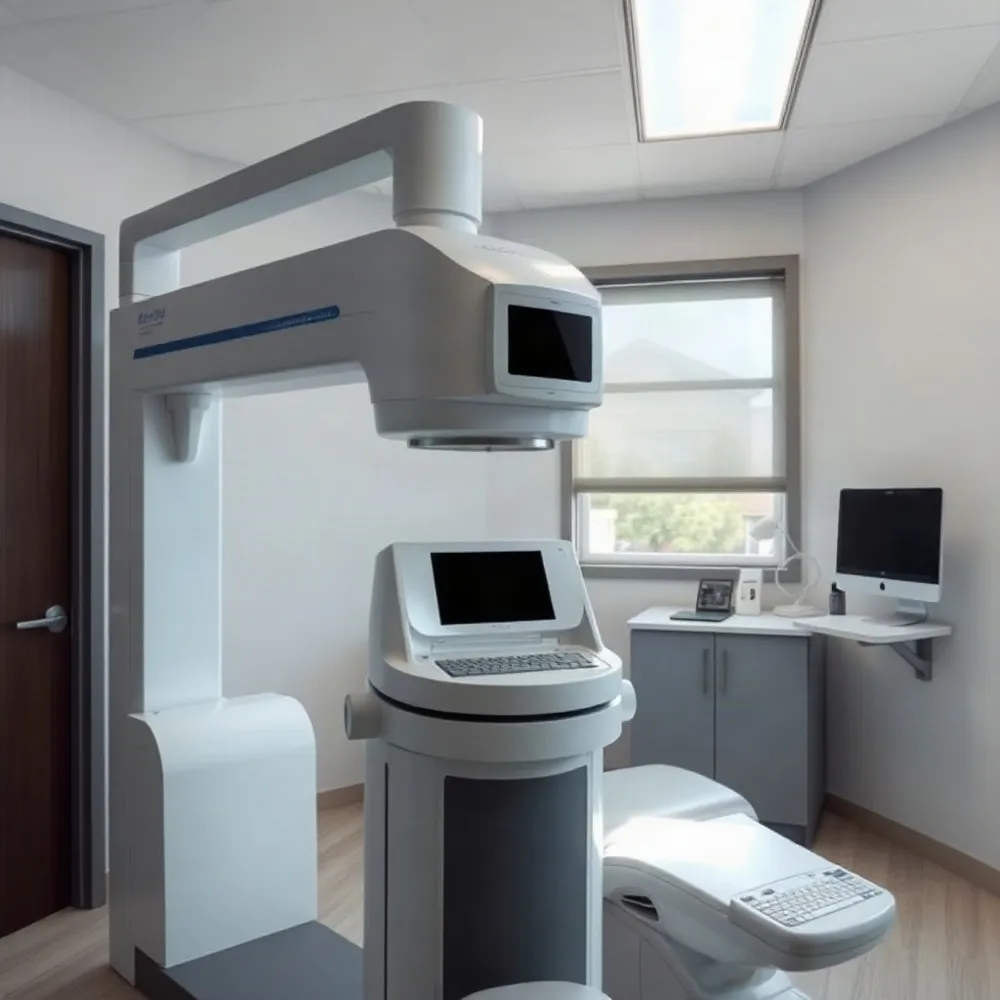 Optical coherence tomography (OCT) is a noninvasive test that uses reflected light to create detailed, cross-sectional images of the eye. It shows the retina’s layers and can also image the optic nerve, without touching the eye. The American Academy of Ophthalmology’s overview of OCT describes how clinicians use these images to examine eye structures.
By measuring tissue thickness and structure, OCT can reveal changes that may not yet affect vision or cause noticeable symptoms. Measurements can help an eye doctor assess conditions such as glaucoma, macular degeneration, and diabetes-related eye disease. Apple Eye Care includes the DRI OCT Triton among its advanced diagnostic tools, as part of a broader optometry practice that also provides comprehensive exams and personalized care.
Repeat scans can help clinicians compare images over time, monitor a known condition, and consider whether findings have changed. The results are one part of an assessment, not a diagnosis on their own. As a general discussion of healthcare technology notes, devices work best when evaluated in the context of patient care and professional judgment (NIH overview of technology in healthcare).
An eye doctor may recommend OCT based on symptoms, examination findings, medical history, or risk factors. It is not necessary for everyone. At Apple Eye Care, the decision to use advanced imaging can be considered alongside the patient’s exam and individual needs.
Conditions OCT Can Help Assess
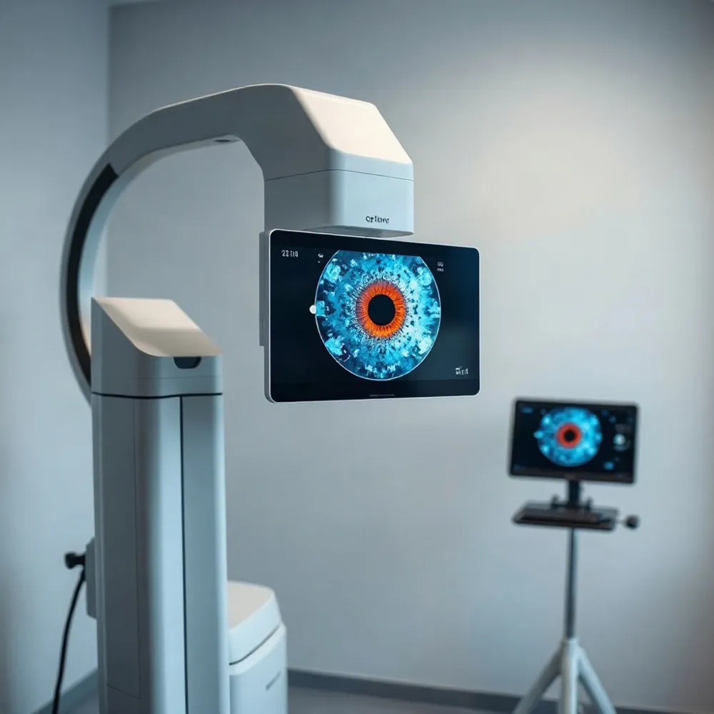 OCT can help clinicians assess changes in the retina, macula, and optic nerve associated with glaucoma and retinal disease. Examples include age-related macular degeneration and diabetic retinopathy, as well as macular edema, macular holes or puckers, and central serous retinopathy. The American Academy of Ophthalmology’s overview of OCT also describes its use in evaluating drusen, vitreous traction, abnormal retinal blood vessels, and blood-vessel blockages.
These images add detail to an eye exam, but they do not determine a diagnosis on their own. An optometrist considers OCT findings alongside symptoms, medical history, and other examination results. At Apple Eye Care, the DRI OCT Triton is part of the practice’s advanced diagnostic technology, supporting assessment within its broader optometry care.
Comparing scans over time can help a clinician identify structural changes and monitor a condition. The findings can inform decisions about follow-up or care, while clinical judgment remains central. This fits the wider role of eye-care technology: appropriate instruments contribute information, and the practitioner interprets it in the context of each patient’s needs, as discussed in research on diagnostic tools in optometry.
The DRI OCT Triton Platform
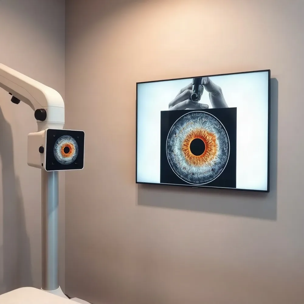 Apple Eye Care identifies the DRI OCT Triton as part of its advanced diagnostic technology. The system combines swept-source optical coherence tomography (OCT) with fundus imaging, giving clinicians different views of eye structures to consider during an examination.
Topcon lists a 1,050-nanometer wavelength and a scan rate of 100,000 A-scans per second for the platform. The manufacturer describes swept-source imaging as designed to visualize deeper structures, including the retina and choroid. These specifications describe the technology, not a guarantee that every structure will be visible in every patient.
Available modes depend on the system configuration. Triton options can include OCT angiography, which shows retinal blood-vessel patterns, and wide-field OCT. Topcon lists fluorescein angiography and fundus autofluorescence for the Triton Plus model only. The DRI OCT Triton product information outlines these configuration differences; it does not establish which optional features are installed at Apple Eye Care.
Images from these modes can add information for a clinician’s assessment and care planning, but they do not determine a diagnosis on their own. Research on healthcare technology similarly stresses assessing tools in the context of patient care and human judgment, rather than assuming newer equipment is automatically better (NIH review of healthcare technology.
At Apple Eye Care, the DRI OCT Triton complements comprehensive optometry services and the clinician’s evaluation. The images are considered alongside the exam and a patient’s individual needs, not as a substitute for professional judgment.
What to Expect During Scanning
What happens during an OCT eye test?
You sit at the scanner and rest your chin, and sometimes your forehead, on supports to help keep your head still. You look at a target while the machine scans each eye with light. Nothing touches your eye, and the test does not use radiation or injections. The scan itself may take about one or two minutes per eye, while an appointment can take roughly five to ten minutes, depending on the exam (Cleveland Clinic’s OCT overview; American Academy of Ophthalmology’s OCT guide). At Apple Eye Care, OCT is part of the practice’s advanced diagnostic services, alongside a comprehensive eye-care approach.
Are there side effects, and can you drive afterward?
OCT is noninvasive and the scan itself generally causes no side effects. Your provider may use dilating drops, but they are not always needed. Drops can leave your eyes sensitive to light and sometimes make vision blurry for a few hours. Ask your provider whether dilation is planned and whether you should arrange a ride; driving is a personal decision based on how clearly and comfortably you can see afterward. Apple Eye Care’s team can explain what to expect for your specific exam. Share any concerns or unexpected symptoms with your eye-care provider.
Reading Results and Planning Care
How are OCT eye scan results interpreted?
A clinician reviews the OCT images for clarity, alignment, and the structures being scanned. They may compare current images with earlier scans to look for changes in tissue thickness or shape. OCT can show retinal layers and help track changes over time, but measurements need to be checked against the images and interpreted alongside symptoms, examination findings, and medical history. A scan or automated measurement alone does not establish a diagnosis. The American Academy of Ophthalmology’s overview of OCT explains how these images can inform diagnosis and treatment planning.
At Apple Eye Care, OCT imaging is part of a wider optometry visit, not a substitute for the clinician’s assessment. The practice offers comprehensive eye exams and specialized care, including glaucoma management, so findings can be considered in the context of your overall eye health.
Is an OCT eye scan worthwhile, and how much does the test cost?
An OCT scan may be useful when an eye-care professional needs a closer look at the retina or optic nerve, or wants to monitor a change. It is not necessary for everyone, and it does not replace a comprehensive eye exam. The Cleveland Clinic’s OCT guide describes OCT as one tool clinicians may use to assess and monitor eye conditions.
Costs vary by practice, scan type, and insurance coverage. Before imaging, ask Apple Eye Care whether the scan is recommended for you and what your plan may cover or the self-pay cost would be.
Technology With a Human Perspective
Advanced imaging can add detail to an eye exam and help clinicians monitor changes over time, but a scan is only one part of the assessment. As the American Academy of Ophthalmology explains, OCT information can support diagnosis and care planning.
At Apple Eye Care, modern diagnostics sit alongside personalized attention and compassionate care. A clinician can discuss findings in the context of your eye health, answer questions, and explain possible next steps without treating a device result as the whole story.